General
Bacteroidaceae family consists of various kinds of bacilli Gram-negative obligately anaerobic Bacteroides, Porphyromonas, Prevotella, Fusobacterium, Wolinella, Leptotrichia, Butyrivibrio, Succinivibrio, Succinimonas, Anaerovibrio, Anaerobiospirillum, Mobiluncus, Selenomonas, Pectinatus, Acetivibrio,
Lachnospira. These bacteria are part of the oral flora, flora of the upper respiratory tract, intestinal and urogenital tract.
The genera Bacteroides, Prevotella and Porphyromas are most important to this family. These bacilli Gram (-) non-spore forming, anaerobes, whether mobile or stationary. They constitute a large part of the normal endogenous flora (oral cavity, respiratory tract, digestive and urogenital) and are now responsible for over 50% of human infections caused by anaerobes.
HISTORY:
In 1920, the first discovery by Bacteroides Castelani and Chalmers.
In 1933, identification on biochemical criteria.
In 1938, the first morphological classification by Provost.
In 1990, genetic classification
CLASSIFICATION:
The latest edition of the 1984 Bergey’s Manual shows the basics of this classification. It is based on the cell morphology, mobility, and the cilia, volatile fatty acids or non-produced cultured and studied by partition chromatography liquid-gas (GLC).
Group 1 = non-mobile bacilli or dliature peritrichous
– Production of butyric acid Fusobacterium
– Production of lactic acid Leptotrichia
– Joint Production of various acids Bacteroides
N = bacilli polar dliature
– Production of butyric acid Butyrivibrio
– Production of succinic and acetic acid Succinivibrio
Succinomonas
– Production of propionic and acetic acid Anaerovibrio
– Production of succinic acid by Wolinella fumarate reduction
Group ni = flagellate bacilli on their concave face
– Fermentative metabolism Selenomonas
– Production of propionic acid and acetic Pectinatus
Group IV = Bacilli flagellates bipolar Anaerobiospirillum

We will limit our study to the most common types in clinical bacteriology: Bacteroides, Porphyromonas, Prevotella, Fusobacterium, Leptotrichia.
The ancient name of the genus Bacteroides was referring to a set phenotype and genotype heterogeneous grouping of many species of bacilli Gram (-) that is used to separate as follows:
 It has recently been established, on the basis of genetic criteria the genus Bacteroides were to be restricted to B.fragilis and related species: B. caccae, B. distasonis, B. eggerthii, B. merdae, B. ovatus, B. stercoris, B. thetaiotaomicron, B. uniformis and B. . vulgatus (formerly known as Bacteroides fragilis group) Other genera were recently separated:
– The kind Porphyromonas combining old and pigmented Bacteroides asaccharolytiques (asaccharolyticus B., B. gingivalis, B. endodontalis).
– The type Prevotella including former non-pigmented Bacteroides and saccarolytiques.
– Finally, other genres have been proposed, most currently with only one species. They concern Gram bacilli (-) uncommon and unclassifiable (Table I).
BACTEROIDES THE GENDER AND RELATED GENRES
Are bacilli Gram (-) of very variable size motionless, the GC% from 35% to 48% (B. fragilis) (5 hypermegas.).
We will deal mainly B. fragilis and content ourselves with some features for other species.
I – BACTEROIDES GENRE:
(Formerly known as Bacteroides fragilis group) Bacteroides fragilis is the type species. Originally called Ristella fragilis, she was isolated in 1898 by Veillon and Zuber in the pus of acute appendicitis then Guillemet in a pulmonary gangrene.
Are bacilli Gram (-) using the hemin as a growth factor. Currently 10 species are distinguished: B. fragilis, B.vulgatus, B. distasonis, B. ovatus, B. thetaiotaomicron, B. uniformis, B. caccae, B. merdae, B. and B. stercoriseggerthii.
A – Habitat and epidemiology:
These are the hosts of the natural cavities of man, especially of the digestive and urogenital tract. They are responsible for the majority of anaerobic infections of endogenous origin.
B – Body type:
They are short bacilli January-March um motionless, non-spore forming, spheroids. A Gram stain, they are often pale with irregular coloration.
C – cropping characters:
They require media supplemented with hemin, incubated in strict anaerobic conditions.
The usual media are represented by the prereduced blood agar and mid-Chalgreen Wilkins.
Culture is slow, at least 24 hours, requiring several days to keep the environments in the oven.
There is then fine colonies, regular and translucent.
Gas production is low in liquid medium.
It is important that the culture is stimulated by the addition of 20% fresh bile.
D – pathogenic natural power:
1. All species are also pathogens:
Anaerobes are responsible for 10% of all bacterial infections and Bacteroides account for approximately a quarter of the pathogenic anaerobes.
During these infections, B. fragilis is the most frequent species (80%). This bacteria has a polysaccharide capsule making it more resistant to phagocytosis.
B. thetaiotaomicron is pathogenic to over 18% of cases.
For cons, the vulgatus species ovatus and distasonis that are most frequent in normal subjects are rarely pathogenic.
2. A break lining and a weakened field are common predisposing factors:
The break contacting a cavity where anaerobic flora is predominant (oral, colonic, vaginal) with a fabric or with the circulation.
The infection starting point is local suppurative thrombophlebitis can cause sepsis and septic metastases.
This mucosal break can be:
– Spontaneous (rupture, perforation)
– Accidental
– Surgical (oral surgery, digestive, gynecological)
Fragile field is frequently involved. Often found as predisposing causes:
– A notion immunodeficiency drug-induced or otherwise,
– Concomitant illness: alcoholism, diabetes, kidney failure, neoplasia,
– A ischémiante condition.
3. Clinical Situations:
Suspected pathogenic role of anaerobic non-spore forming before a foul pus, located near a mucosal tissue necrosis with … and when previous treatment with aminoglycosides or beta-lactam antibiotics was ineffective.
The tables are in the form:
– Septicemia: Bacteroides being most frequently involved as Fusobacterium,
– Stomatological infections,
– ENT infections: otitis, sinusitis, thrombophlebitis,
– Lung infections: pneumonia, lung abscess, pleurisy (P. melaninogenica)
– Abdominal infections: appendicitis, peritonitis, intra-abdominal abscesses, perianal infections (B.fragilis)
– Gynecological infections: post-abortion, post-partum, salpingitis, pelvic infections following gynecologic surgery,
– Infections of the central nervous system: brain abscess (B. fragilis), sub dural empyema, meningitis,
– Other locations: skin, bone and joint …
E – Experimental pathogenicity:
Some strains can cause abscesses accompanied by cachexia and resulting in the death of the animal within 10 to 20 days.
F – Diagnosis:
1. Bacteriological Diagnosis:
Isolation poses no particular problem provided you have the appropriate backgrounds and achieve a good anaerobic conditions.
The systematic practice of specific samples for anaerobic (blood cultures, wound infection, pleural fluid, peritoneal or other) currently allows to put often highlighted.
Growth is slow and sometimes request 4 to 5 days. In samples where Gram-negative facultative anaerobic bacilli can make the difficult isolation, use a selective medium supplemented with aminoglycosides (kanamycin, gentamicin).
The identification is based primarily on a favored growth in the presence of bile, indole research and study of the fermentation of sugars. Carbohydrates are widely used as the fermentative pathway.
Bacteroides species are very similar, so they are all sucrose (+), lactose (+), maltose (+) xylose (+), mannose (+).
Other characters to be considered are: esculin (+), gelatinase (-), milk (-), H2S (-).
The distinction is made on indole, cellobiose, mannitol, arabinose, salicin, glucose, trehalose, rhamnose (Table II).The existence of miniaturized galleries ready to use in this case allows easy diagnosis.
The gas chromatography allows in difficult cases to distinguish between Bacteroides and Fusobacterium, but it is not usually necessary to differentiate Bacteroides species. It may be useful to separate Fusobacterium species that can grow in the presence of bile .

2. Rapid Diagnosis:
Identification by immunofluorescence with anti-B serum capsule fragilis is possible either directly on the pathological product is from a liquid medium enrichment.
II – TYPES RELATED:
A – pigmented species (Prevotella, Porphyromonas):
The pigment grows on medium supplemented with hemin, lives K.3 (menadione) and containing 5% total or lacquered sheep blood. It is within 3 to 21 days, however, it may not develop.
This group includes 8 species, the best known are P. melaninogenica and P. . asaccharolytica Morphologically it is often coccobacilli Gram (-) stationary non-spore forming.
The main characters of identification of these species are summarized in Table III.

1. Cropping characters:
Colonies appear very fine and only after 3 to 4 days, there may be a UV light by fluorescence. Then they grow, pigmented and surround themselves with a slight hemolysis halo.
2. pathogenic natural power:
In isolation, these bacteria are not pathogenic; but their presence potentiates the virulence associated bacteria.
Melaninogenica P., P. gingivalis, P. endodontalis, P. oris, P. buccae, P. intermedia are preferentially isolated from oral samples or pleuropulmonary.
Asaccharolytica P., and P. bivia meet especially in the abdominal and genital infections.
B – saccharolytic Cash and bile-sensitive:
There are 3 groups:
a / pentose-fermenting species: P. oris, P. buccae, P. zoogleoformans isolated in dental and lung infections.
bl Species fermenting pentoses P. oralis, P. buccalis, P. veroralis.
cl Species saccharolytic and proteolytic; P. bivia, P. saying.
The identification characters of the main species are summarized in Table IV.

C – Bacteroides non saccharolytic:
B. Putredinis
It can be isolated from intestinal infections (appendicitis) and does not require a growth factor.
B. ureolyticus (anc. B. corrodens)
It has a typical appearance culture colonies flat, shiny, digging the culture medium (possible confusion withEikenella corrodens)
It has few positive characters (do not ferment sugar).
It is isolated in oral or pleuropulmonary infections.
B. pneumosintes
This is a very small bacillus, colonies are visible only magnifying glass, the culture is slow. It ferments no sugar and can be isolated from nasopharyngeal samples.
The main characters of identification are summarized in Table V. They are esculin (-).

III – TREATMENT OF INFECTIONS AND SENSITIVITY BACTEROIDESANTIBOTIQUES:
Treating Gram anaerobic infection (-) based on two types of action to be taken together.
A – Local treatment:
D include the evacuation of pus (puncture, surgical excision), excision of necrotic tissue and tissue exposure to oxygen or hyperbaric chamber.
B – General treatment: antibiotics:
Anaerobic are constantly resistant to aminoglycosides, these antibiotics can not spend the bacterial membrane.
The problem is dominated by Bacteroides fragilis is the species most common and most durable.
Penicillin G, the longest used in anaerobic infection, is ineffective B. fragilis, which is resistant to many beta-lactam antibiotics through a TEM beta-lactamase. Beta-lactams are generally effective: ureidopenicillins, associations with a beta-lactamase inhibitor, imipenem.
Among the cephalosporins, cefoxitin is the most effective; 3rd generation cephalosporins are not most active, with the exception of cefotetan. Bacteroides fragilis is resistant to tetracyclines in 60% of cases and 10% in the macrolide.This resistance is plasmid or chromosomal origin.
Chloramphenicol is very efficient (very few strains of B. fragilis resistant) and has the advantage of a good pass in the central nervous system, but its toxic effects limit signs.
Clindamycin offers the same advantages and similar disadvantages.
Metronidazole has a specifically anti-anaerobic action and there is virtually no encounter resistance amongBacteroides to this antibiotic although it is used for a long time. The finding of a resistant strain in vitro métronidazone must discuss identification.
Metronidazole act as specific metabolite of fermentative way by operating irreversibly alternative and as a terminal electron acceptor in a phosphorylation reaction at the substrate. Now the way is fermentadve capital among anaerobes. We showed that the metronidazole is reduced ferredoxin or by metabolic processes associated ferredoxin in susceptible anaerobic bacteria. This reduction is important for the antibacterial action:
– By decreasing the intracellular concentration of non-reduced compound, creating a gradient which allows the entry of metronidazole in the cell,
– Giving birth to responsible derived from the destruction of sensitive bacteria.
In practice for an infection to B. fragilis, use a bi-antibiotic which is an antibiotic metronidazole and the other will be adapted according to the antibiogram. Always think that these infections are often polymicrobial (Bacteroides, Fusobacterium, Clostridium, facultative anaerobic bacteria …), piperacillin then represents an interesting choice.
Other Bacteroides species are more rarely producing beta-lactamase than is B. fragilis, they are generally sensitive to penicillins and cephalosporins.
FUSOBACTERIUM GENRE
It is bacilli Gram (-) anaerobes, very variable in size, non-spore forming, sometimes moving (ciliature peritrichous) and characterized by a large polymorphism. Their GC varies between 26% and 34 mol. They do not require haemin for growth.
They produce a significant amount of butyrate with a characteristic odor.
Ten species are described in the latest edition of Bergey’s Manual: F. gonidiaformans, F. mortiferum, F. naviform, F. necrogenic, F. necrophorum, F. nucleatum, F. perfoetens, F. prausnitzii, Russii F., and F. varium.
Recently new species have been described, they are present in the oral cavity, it is F. periodonticum, F. alocis, F.and F. Suici simiae.
I – Fusobacterium nucleatum:
Described in 1896 by Vincent in angina and phagedenic ulcer (in association with spirochetes). Culture is performed by Veillon and Zuber in 1898 (former names: Fusiformis fusiformis, Sphaerophorus fusiformis).
A – Habitat:
This is a commensal bacterium of the oral cavity and the respiratory tract, it can also be isolated in the digestive tract or genital.

B – Body type:
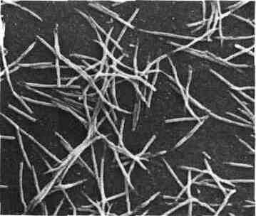
These are bacilli Gram (-) with tapered ends where an aspect spindle (shuttle) lying, very variable in size (2 nm to 10 microns long).
Fusobacterium is capable of being transformed in filamentous form with terminal or median indentations which could lead to free spheroplasts January-March um diameter. Metachromatic granules may be present. This is an immobile bacteria.
C – cropping characters:
It is a strict anaerobic germ, but some strains can tolerate up to 6% O2 (microaerophilic).
Pathogenic strains are more demanding than saprophytic strains and sérophiles are required. Colonies can be in three aspects: smooth, mottled or breadcrumbs. Broth large flakes training is observed (appearance of bread). This species is little or no gasifier.
D – biochemical characters:
Attack of carbohydrates is low.
The important characteristics are:
– Gelatinase (-).
– Milk slowly coagulated.
– Indole (+).
– GLC: butyrate peak.
E – pathogen natural power:
Fusobacterium nucleatum is the species most frequently isolated from Fusobacterium.
He is responsible for Vincent’s angina is ulceration of tonsils covered with a pseudo-membranous grayish coating.Bacteriological diagnosis is obvious after Gram staining: presence of fuso-spirillaire association.
This seed is predominant at the pulmonary tract where it can be at the origin of purulent pleuritis, lung abscess.
In smaller proportions he is responsible for various infections:
– Oral: stomatitis, pulpitis, dental cellulitis,
– Disorders: liver abscesses, peritonitis, colitis,
– Genital,
– Meningitis, adenitis, …
As with Bacteroides, we note that a particular terrain favors the occurrence of infections or secondary infections due to this organism.
There is no production of toxin or hemolysin.
F – Diagnosis:
It will be discussed by the morphology and confirmed by the study of the biochemical characteristics (see Table VI).Three subspecies have been described differing only in their electrophoretic profile and GC%.

II – FUSOBACTERIUM necrophorum:
This species has been described in the literature as thirty denominations all synonyms Sphaerophorus necrophorus, Sphaerophorus funduliformis, botch necrophorum aids …
This is one of the few non-spore forming anaerobes whose pathogenicity is due to toxic factors.
A – Habitat:
II is in natural cavities: oral cavity essentially vagina.
B – Body type:
This is a bacillus Gram (-), immobile, highly polymorphic: short forms ovoid to clear center, long filamentous forms with bulges spheroplasts.
Recently described F. pseudonecrophorum different from F. necrophorum by the absence of lipase and beta hemolysis.
C – cropping characters:
This is a strictly anaerobic bacterium. The optimum pH is slightly basic development (7.5-7.8).
Colonies are gray or yellow, convex, 2 to 3 mm in diameter with a raised center (egg appearance on the flat) and sometimes a little hemolysis halo.
Together there is a foul smell.
Broth, the culture is very flaky and gasifier.
It is a non sérophile species.
D – biochemical characters:
The main characteristics to be considered are:
– Glucose, maltose, galactose (+),
– Lactose, trehalose, raffinose (+ or -)
– Indole (+),
– Gelatinase (-)
– Slowly coagulated milk,
– GLC: significant production of butyrate; Propionate and acetate are also present in lower amounts.
E – pathogen natural power:
F. necrophorum was responsible for necrotic acute angina followed by severe sepsis fatalities before penicillin. It may also be responsible for gynecological infections, otitis, mastoiditis, brain abscess, digestive infections: liver abscess, appendicitis, peritonitis, septicemia. However, these infections are more rare now since the use of penicillin.
The combination of angina and pulmonary infarction due to F. necrophorum is Lemierre syndrome.
F – Experimental pathogenicity:
The intravenous injection to rabbits or guinea pig of a culture of 48 h in liquid medium results in a fatal septicemia.
Pathogenicity is due to a hemolysin and hemagglutinin.
III – OTHER SPECIES FUSOBACTERIUM:
A – Fusobacterium mortiferum:
Formerly known as F. freundii or Sphaerophorus mortiferus is a host of the oral cavity and gastrointestinal tract. It is isolated in many intestinal abscesses, pleurisy etc …
It ferments many sugars. The differential diagnosis can arise with some Bacteroides, in this case the GC is useful.
It is characterized by its resistance to macrolides and rifampicin.
B – Fusobacterium necrogenic:
II can be isolated in the stool.
C – Fusabacterium varium:
II can be isolated from the oral cavity, rarely considered pathogenic.
IV – TREATMENT AND ANTIBIOTIC SENSITIVITY OF FUSOBACTERIUM GENRE:
In sepsis, surgery to remove the front door is essential.
Overall species of the genus Fusobacterium are characterized by their high sensitivity to beta-lactams, macrolides, chloramphenicol and usually antibiotics active against anaerobes. However, strains with a beta-lactamase can be isolated.
LEPTOTRICHlA GENRE
It was described in 1879 by Trevisan. This genus has long been confused with Fusobacterium nucleatum.
I – GENERAL CHARACTERISTICS:
They are free or little curved bacilli from 5 to 15 microns of 1 (im with tapered or rounded ends, motionless. One can find some filamentous forms and / or spheroplasts.
GC = 34%.
GLC: predominant peak of lactic acid, acetic acid producing low, never produce butyric acid.
One species is recognized: Leptotrichia buccalis.
II – HABITAT – EPIDEMIOLOGY:
It is a commensal organism of the mouth, sometimes isolated from the digestive tract or vaginal.
It can be found as opportunistic pathogen in immunocompromised.
III – CHARACTER BACTERIOLOGICAL-IDENTIFICATION:
A – cropping characters:
The colonies appear only after 4-5 days oven, together with a putrid gassing.
These colonies are irregular (shaped jellyfish).
B – biochemical characters:
Glucose (+), lactose (+), sucrose (+), maltose (+), raffinose (+).
Indole (-), H ^ S (-) Catalase (-), nitrate reductase (-), gelatinase (-), mannitol (-).
C – Differential diagnosis:
D can arise with F. nucleatum.
D – Treatment:
This germ is sensitive to beta-lactams, macrolides, chloramphenicol.
You must be logged in to post a comment.