Borrelia are responsible for borreliosis, usually called recurrent fevers. These are diseases transmitted by blood-sucking arthropods. According to the arthropod vector is conventionally divided into two groups of borreliosis:
– The borreliosis transmitted by lice,
– The tick-borne borreliosis.
This bacterial genus experiencing a revival since the isolation of
B. burgdorferi, the agent of Lyme disease called and related conditions.

HISTORY:
A – Relapsing fever:
They are known since Hippocrates. This is the nineteenth century that were individualized clinically recurrent fevers, and we isolated a pathogen: sanguicole a spirochete.
In the early twentieth century were highlighted vectors Agents: First lice, ticks then. In 1907, spirochetes responsible for these fevers were named Borrelia in honor of the French bacteriologist A. Borrel.
B – Lyme disease and related conditions:
B. burgdorferi is responsible for several conditions:
– Erythema chronicum are migrating or erythema Lipschutz (ECM).
– Said Lyme Disease.
– Chronic or relapsing disease Acrodermatitis Pick-Herxheimer (ACA).
– Meningoencephalitis radiculo-neuritis Garin-Bujadoux-Bannwarth.
– Lymphadenosis benigna cutis (LABC): Benin lymphocytoma skin.
The outstanding historical stages are:
1909 first description of ECM Afzelius.
1913 establishment by Lipschiitz criteria ECM and vector role of the genus Ixodes ticks.
1920-1945: first descriptions of other ailments.
1955: experimental demonstration of the infectious etiology.
1970 description of the first case of ECM in the state of Wisconsin, USA
1975: Steere survey among residents of Lyme localities suffering from rheumatoid arthritis adjunct of an ECM
1978: Highlighting the role of the vector Ixodes dammini by Steere et al.
1982: isolation of the pathogen in ticks collected in Shelter Island in the state of New York by Burgdorfer; similar discovery in Switzerland Ixodes ricinus.
1983 isolation of the pathogen in human samples by Steere et al.
I – HABITAT – EPIDEMIOLOGY:
Borrelia belong to the order Spirochaetales and family Spirochaetaceoe.
The genus Borrelia includes twenty species whose main epidemiological characteristics are listed in Table I.

A – Vectors:
Their distribution determines the geographic range of borreliosis.
All species of Borrelia are transmitted by arthropods. These vectors are either blood-sucking lice or ticks.
Transmission of B. burgdorferi is mainly done by arthropods: mites (family Ixodidae) and to a lesser extent by insects (family Culicidae and Tabanidae).
1. Lice:
These are Anoplura insects. The only interesting species is Pediculus humanus medical pathology.
All stages (larvae, nymphs, adults) are bloodsucking. The louse is contaminated by biting an ill person. The spirochete Borrelia recurrentis is involved.
In the louse, B. recurrentis only found in the hemolymph and it invades or salivary glands or the intestine; so the bite is never infective to humans and contamination is possible only in the crash of the louse (delousing, scratching).Germs can then actively penetrate the skin and mucous membranes.
2. Ticks:
There is a specificity between the vector species and species of Borrelia infective.
These are:
– Either soft ticks mites: the omithodores or Ornithodoros.
The omithodore becomes infected by biting an infected animal and ingested Borrelia disseminate in all its tissues.The infection can be transmitted to a healthy body by stitching the coxal fluid released at the end of blood meal
– Or Ixodidae mites: Ixodes.
Borrelia burgdorferi is located at the middle gut of the tick. Transmission to humans by the tick during its blood meal;there is probably also a transmission by saliva and regurgitation of intestinal secretions.
Ixodes ricinus seems to be, in Europe, the main tick vector. In the USSR, I. persulcatus has been incriminated as a vector of the disease.
B – Tanks:
B. burgdorferi is the only species capable of infecting mammals and birds; many wild animal species and are potential reservoirs of B. burgdorferi. In North America, the main reservoirs are mice (Peromyscus) and deer. In Europe, the tanks are voles, field mice, deer, foxes and wild boars. Pets peuvtent also serve as a reservoir.

C – Distribution:
It is generally summarized globally in Table 1, which shows for each major type of borreliosis (s) agent (s) pathogen (s), their (s) vector (s), their (s) tank (s ), the type of contamination and the infected areas.
Figure 1 shows the range of ticks in Europe. There is a narrow specificity between the vector parasite (/. Ricinus)and the agent of Lyme borreliosis.
II – NATURAL PATHOGENICITY:
A – The human relapsing fever:
Schematically the typical clinical picture associates after a silent invasion phase of one week or more:
– A fever of 39 to 40 ° C to sudden onset;
– Diffuse pain syndrome;
– A hepatosplenic with jaundice syndrome, digestive disorders, moderate hepatomegaly, splenomegaly net;
– A neuro-meningeal syndrome with frequent dizziness, intense headache. A lumbar puncture cerebrospinal fluid is clear with lymphocytosis. Centrifugation can reveal Borreha in the pellet;
– A mucocutaneous syndrome with dry skin (by difference with malaria), a wet tongue covered with a characteristic mustard plaster, often rash, epistaxis.
This table takes a few days and then there is an abrupt defervescence accompanied by sweat and urine crisis. This afebrile phase lasts one week and then the same symptoms reappear (recurrence). The number of repeats varies from 3 to 10 on average according to the species Borrelia.
We distinguish five main types of recurrent fevers which have their own clinical features (Table I).
B – diseases caused by B. burgdorferi:
Schematically are three main evolutionary stages of the disease:
1. Primary stage:
It is characterized by a typical dermatological lesion YErythema Chronicum are migrating (ECM) or erythema Lipschiitz. This comes days after the tick bite in the form of a rash papular gradual extension and centered by the bite. It may be accompanied by fever, headache, fatigue, lymphadenopathy, arthralgia. Sometimes the picture is more alarming (neurological damage, liver, joints).
2. Secondary Stage:
The secondary stage manifestations may occur weeks or months after the ECM They can be isolated or associated with each other.
a / Skin manifestations:
Lesions resemble the original ECM, but are generally smaller.
b / Cardiac Events:
It is usually impaired atrioventricular conduction, but myocarditis and pericarditis may be observed. These events usually lend itself within a few days to a few weeks.
c / Joint Events:
They are characterized by brief episodes of asymmetric trace arthritis most commonly affects the large joints (knee).
d / Neurologic Manifestations:
They can start the clinical picture outside the occurrence of an ECM combining variably lymphocytic meningitis, cranial neuritis (facial paralysis essentially) and often asymmetric and very painful radiculitis meningoencephalitis.
This table is close to that of the meningo-radiculitis described by Garin, Bujadoux and Bannwarth.
Some tables are dominated by central events: cerebral accident going tem, demyelinating encephalitis, comatose major disorders.
3. Tertiary Stage:
Late manifestations are characterized by their chronicity.
a / Joint Events
They come in the form of trace inflammatory arthritis of large joints, relapsing for years and that may result in osteochondral lesions.
b / Acrodermatitis chronica atrophicans (ACA) or Pick-Herxheimer disease
Affection especially women, ACA is a chronic skin disease of progressive evolution, resulting in spontaneous inflammatory atrophy of the integument.
It can be complicated benign skin tumors Benin lymphocytoma skin type, malignancies (lymphoma B).
III – PATHOPHYSIOLOGY:
A. Relapsing fever:
Few facts are known. The important point is the recurrence mechanism: during his time in the human body, the spirochete present successive antigenic variations responsible for the absence of specific antibody production, each of these antigenic variations resulting recurrence. In B. hermsii, these variations are encoded by linear plasmids.
B. disorders due to B. burgdorferi:
B. burgdorferi against, shows no antigenic shift.
Antigens or potential virulence factors, specifically responsible for the expression of the pathogenicity are poorly understood at present. Both plasmids and two surface proteins, OspA and OspB of 31 kDa and 34 kDa respectively, appear to be associated with virulence. In addition, a “endotoxin-like” activity due to lipopolysaccharide was highlighted. Significant correlations have been established with certain HLA groups (DR2).
The study of DNA and of European and American strains structural proteins revealed the existence of different antigenic proteins between these two groups of strains. This concept could explain the existence of two types of clinical presentations. It is currently moving towards the recognition of the existence of two subspecies of B.burgdorferi.
IV – BACTERIOLOGICAL DIAGNOSIS:
A – disorders due to B. burgdorferi:
1. Samples:
The pathogen can be sought in the cerebrospinal fluid, skin lesions, peripheral blood; but it is an extremely fragile germ.
2. Direct examination:
The study can be done using a dark-field microscopy or after staining (Giemsa more often, or film sensitive realization).
Will examine the pellets centnfugation the cerebrospinal fluid, tissue sections.
3. Experimental pathogenicity:
It is not used for diagnostic purposes, but is at present a model for the study of the pathogenesis of these infections.The most commonly used animals are rabbits, guinea pigs and hamsters.
4. Culture:
For the cultivation of B. burgdorferi are currently used the liquid medium Barbour-Stoener and modified Kelly (BSK II Barbour et al. 1984) were incubated at 33 ° C.
Inoculation is on the surface and future growth is sought in microaerophilic by taking every week for several months.
The complex composition of this medium BSK II uses chemical and microbiological products, whose quality and brand are fundamental to the success of crops.
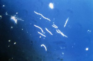
5. Morphology – Structure:
Spirochetes are 10 to 30 microns long and 0.2 to 0.3 microns in diameter. They have 5-7 turns on average, slightly irregular and tightened, they are movable in rotation and translational movement associated.
Structurally, they are surrounded by a flexible outer envelope. Cytoplasm surrounded by a membrane containing parieto-cytoplasmic peptidoglycan.
6. Serological diagnosis:
Culture is difficult and late results. Laboratory diagnosis of these disorders is currently mainly HIV. B. burgdorferi is the only species of Borrelia why serological diagnosis is possible.
The interpretation of this serology is the subject of much discussion.
Two techniques are commonly used: the IIF and ELISA.
– The immunofluorescence (IIF), the most used technique in Europe is conducted using an antigen consisting of a culture of B. burgdorferi affixed blade by a conventional technique similar to ETS.
– The ELISA described by Russell et al. and by Magnarelli et al. in 1984, using a soluble antigen obtained from a culture treated with ultrasound in the first case and a suspension of inactivated spirochetes in the second. The revelation of the antigen-antibody reactions are made with anti-human immunoglobulin sera labeled with alkaline phosphatase or with peroxidase.
Positivity thresholds vary between laboratories, but can be considered significant:
– In IFI IgG 1/64 th (1/256 some authors)
IgM 1732nd
– In ELISA IgG 1/200 th
False positive cross-reactions occur with Treponema pallidum and rarely with Leptospira. In these conditions, the very different clinical context makes the differential diagnosis. Further note that in some patients with positive serology MNI observed 30% false positive results.
A better approach serological diagnosis is provided by the study of the serum and / or CSF by Western blot. This technique of immunoblotting of revealing through an immunoperoxidase reaction, antibodies directed against theBorrelia antigens separated by polyacrylamide gel electrophoresis and transferred to nitrocellulose. IgM and IgG antibodies detected in the serum of patients vary depending on the stage of the disease. We must assess the number of antigens and revealed their relative importance to give an interpretation. The Western blot is more sensitive than the IFIs and helps to improve the diagnosis, including neurological forms.
Other tests can be used (passive hemagglutination and immunocapture IgM).
It is interesting to study the positivity rate and the evolution of IgG and IgM antibodies in the different conditions (Table II and Figure 2). In case of complex ECM, antibodies remain high away from the sting while for ECM “simple” one negativity was observed after a while. Can we infer that a high and persistent levels must fears the occurrence of complications?
Table II shows that the serodiagnosis positivity rate is much higher in case of complications during an ECM. 2 shows elevated IgM in the ECM (early phase) and then decreasing in neurological and cardiac involvement (late phase).Conversely IgG is low for the ECM and grow to be realized when arthritis sets in. Is it a simple chronological evolution or is it a reflection of disease severity?


7. New diagnostic tools:
The methods of genotypic detection by in vitro DNA amplification (polymerase chain reaction) appear to be a promising tool that can effectively replace culture for the diagnosis of these infections from a variety of organic products. Progress remains to be done to popularize these methods.
B – Relapsing fever:
For these borreliosis, diagnosis is essentially microscopic.
It can be effectively supported by experimental pathogenicity. Intramuscularly inoculated by intraperitoneal or blood or infected tissue homogenate. The most sensitive animal is Swiss mice and newborn rat. One can also use the guinea pig and rabbit. If the sample is positive, the animal is a disease similar to that of man, and this in a period of less than 15 days. Blood smears will be realized every 3 days to search after Giemsa spirochetes.
There are no status. The clinical and epidemiological confrontation is dominating.
Regarding the culture, the medium of the Kelly or BSK II medium can be used, but do not allow the growth of certain species: B. hermsii, B. parkeri, B. turicatae, B. hispanica, B. recurrentis.
V – THERAPY – PROPHYLAXIS:
Borrelia are very sensitive to antibiotics germs. Preferably used for uncomplicated forms a beta-lactam antibiotic (ampicillin, amoxicillin, 2 g / day for 10 days) or a macrolide or a cyclin.
Ceftriaxone (IV, 2 g / day 15 days to 3 weeks) is recommended and seems more effective in treating complicated forms, especially neurological.
In infections with B. burgdorferi, it seems that early treatment significantly reduces the risk of developing late onset and shortens the duration of evolution of ECM as well as the occurrence of cardiac and joint damage.
Be wary of too aggressive treatment may lead to Jarisch-Herxheimer.
Regarding prevention, eradication of vectors is difficult to achieve; individual prophylactic measures are effective. It does not exist at present vaccine.
You must be logged in to post a comment.