HISTORY:
In 1913 a mobile bacillus Vibrio-like, was made responsible for the epizootic bovine abortions tasks. This bacillus was designated as Vibrio fetus by Smith and Taylor (USA) in 1919. A French bacteriologist Vinzent, reported in 1949 the first case of human infection with Vibrio fetus.
In 1963, through the study of G + C% of DNA, Sebald and Véron showed that these bacteria are different from Vibrio(G + C% = 40 to 52) and form a new genus, Campylobacter (G + C% = 30-38).
In recent decades the conditions of isolation of Campylobacter have been improved and the role of these bacteria as agents of enteritis has been demonstrated.
I – DEFINITION:

Campylobacter are Gram-negative bacilli characterized by:
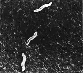
– Morphology. Bacilli purposes, from 0.5 to 5 | J, m long, curved comma-shaped, S-shaped, the “gull-flight” or helical shape to the long forms.
-. The mobility is intense due to monotriche polar cilia. It is classically described as a “gnat flying.” The long forms may be flagellated at both ends.
microaerophilic their respiratory metabolism.
oxidase reaction (+).
a G + C% of the DNA comprised between 30 and 38%.
II – CLASSIFICATION:
Catalase is used to divide the genus Campylobacter in two groups.
A – catalase negative group:
The species that make up this group are not pathogenic for humans.
C. sputorum is divided into three subspecies:
– Subsp sputorum, found in normal oral flora..
– Subsp bubulus, found in preputial secretions of the bull..
– Subsp mucosalis isolated intestinal adenomas pork..
B – positive catalase group:
In this group there are:
– C fe tus, with two subspecies: C. . fetus subsp fetus responsible for septicemia in the immunocompromised, it is also pathogenic to cattle and sheep, C. fetus subsp. venerealis is responsible for abortions and livestock sterility.
– C jejuni is now recognized as a common bacterium responsible for diarrhea in humans.
– C coli is close to C. jejuni and has the same pathogenicity.
C – Other species:
Other recently recognized species are attached to the genus Campylobacter:
– C. laridis is common among gulls,
– C.fecalis found in cattle, but not humans,
– C. hyointestinalis gives enteritis in pigs
– C. cinaedi and C.fennelliae were found in the rectum homosexuals,
– C. cryaerophila and C. nitrofigilis were found in the environment.
– C. pylori or C. Pyloridis often associated with damage to the lining of the stomach, is now classified in the genusHelicobacter.
III – HABITAT AND EPIDEMIOLOGY:
Campylobacter are bacteria found in the digestive tracts of animals, especially poultry, sheep and swine. Pets (dogs and cats) have been identified as Campylobacter vectors.
Contamination of humans by digestive track. Cases of campylobacteriosis are often sporadic. The contaminated water or dairy products were at the origin of epidemics.
IV – PATHOPHYSIOLOGY:
Because of their high mobility Campylobacter are able to penetrate the mucus. They can enter the enterocytes.Invasiveness of the bacteria results in the presence of leukocytes and blood in the stool of patients.
Toxin has been demonstrated in strains of C. jejuni. One of them is closely related to cholera toxin. Another toxin would cytotoxic activity. Their exact role is still unclear.
V – PATHOGENICITY:
A – Human health:
– C fetus subsp fetus is responsible for septicemia digestive start occurring in pregnant women or in patients with underlying disease.. cirrhosis, blood disease, AIDS etc … Localized infections (cardiac, meningitis, joint) also been described.
– C jejuni and C. al. They are much more often encountered. They are due to enteritis which are more common in children living in poor hygienic conditions. After incubation for 1 to 3 days, febrile diarrhea occurs sometimes with blood in the stool. Abdominal pain and vomiting are common. Healing occurs spontaneously in about a week.Bloodstream infections are infrequent. Some infectious complications (appendicitis, cholecystitis, peritonitis) or post-infectious (arthritis) have been reported.
B – For the animal:
– C fetus subsp. fetus is found as a commensal in the intestine of cattle, sheep, pigs and poultry. From the gut bacteria can have a blood-borne; the infection can be localized to the genital tract and cause “sporadic abortions.”
– C fetus subsp. venerealis only found in animals in which it causes the “enzootic sterility” of venereal origin.
VI – BACTERIOLOGICAL CHARACTERS:
A – Campylobacter are microaerophilic:
They need for their growth an atmosphere having a reduced oxygen content, but they have a respiratory metabolism and do not grow anaerobically.
The most favorable to their growth gaseous mixture contains 5% oxygen, 10% CO2 and 85% nitrogen. It is most easily achieved by using a suitable generator placed in a jar for anaerobes.
B – Culture media:
Campylobacter is developing on-Columbia agar supplemented with 5% blood. They also develop on Brucella agar and in Mueller-Hinton medium.
The growth in semi-agar (0.15% agar) as thioglycolate broth is better than in liquid medium.
Selective media to search for Campylobacter in feces have been developed. They contain a mixture of antibiotics inhibit most bacteria coproflore:
– Mid Butzier: bacitracin, novobiocin, cycloheximide, cefazolin, colistin.
– Skirrow: vancomycin, trimethoprim, polymyxin.
– Mid Blaser: vancomycin, trimethoprim, polymyxin, cephalothin, amphotericin.
Campylobacter grow at 37 ° C, but an incubation temperature of 42 ° C promotes the growth of C. jejuni.
C – Aspect of colonies:
On agar colonies appear within 2 to 4 days. They are 1 to 2 mm in diameter. They are flat, gray and translucent. They may have an irregular edge. When the agar is not completely dry, the colonies can be spread. They sometimes have a mucoid appearance.
When the colonies are elderly, bacterial bodies become coccoid. This form of degeneration is irreversible.
D – biochemical characters:
– All Campylobacter reduce nitrate to nitrite and are oxidase (+).
– Species of medical interest all have a catalase.
– They do not hydrolyze sugars.
– C jejuni is distinguished by its ability to hydrolyze hippurate.
The characters that distinguish the major Campylobacter species encountered in human medicine are shown in the table below:

VII – RESEARCH C. JEJUNI IN THE SADDLE:
– It should be performed on freshly issued stool or stored at 4 ° C.
– The direct microscopic examination phase contrast or black background with a drop of fecal suspension is a critical time. It often helps to recognize bacilli with the characteristic mobility.
– The stool microscopy after staining with methylene blue can show significant amount of polynuclear who witnessed the invasiveness of the bacteria.
– The culture is generally made by inoculating selective media described above. Another possibility is to filter a stool suspension onto a filter of 0.65 [t, which is then applied directly to the agar and retained one hours medium.
VIII – ANTIBIOTIC TREATMENT:
Or septicemic forms other than digestive campylobacteriosis warrant antibiotics systemically.
For Campylobacter enteritis whose recovery is spontaneous, the need for antibiotic treatment is discussed. The antibiotic of choice is erythromycin which shortens the length of the digestive portage.
Campylobacter are sensitive to aminoglycosides, tetracycline, chloramphenicol, and the combination amoxicillin-clavulanic acid. Sensitivity to ampicillin is inconstant. They are resistant to colistin, cotrimoxazole, rifampin and cephalothin.
You must be logged in to post a comment.