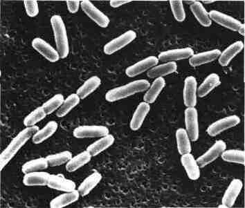
Isolated for the first time by Escherich in 1885, is the Escherichia coli bacterial species that has been most studied by fundamentalists for physiology and genetics work. This bacterium has long been known as commensal and pathogenic digestive tube to the urinary tract. In recent decades, the role of certain categories of E. coli in diarrheal syndromes was clarified and the mechanisms of the pathogenicity were analyzed.
I – HABITAT:
E. coli is a commensal species of the gastrointestinal tract of humans and animals.
In the intestine, E. coli is aerobic species quantitatively the most important, present from 10 ^ 109 bacterial cells per gram of stool. This bacterial population is only about 1% o of the anaerobic (see box on the flora of the digestive tract).
Search E. coli in the water supply (colimétrie) is done to assess its potability. The presence of E. coli in water is the indicator of recent faecal contamination and makes it unfit for consumption.
II – PATHOGENICITY:
A – pathogenicity for man:
1. Intestinal infections:
The presence of diarrhea in E. coli has been known since 1940.
These diarrhea are caused by specific strains of serotypes that cause either sporadic cases or small outbreaks.
The different clinical syndromes are due to E. various coli which we will specify later the support virulence. Now recognized four types of strains causing diarrhea:
a / enteropathogenic strains or “Entero-Pathogenic E.coli” (EPEC):
They were responsible, in the 50s, severe toxicosis or infantile diarrhea occurring in outbreaks in nurseries and maternity. These strains also called E. GEI coli (infantile gastroenteritis) are rarely encountered today they are then isolated sporadic cases. They belong to specific serotypes: YES, 026, 055, 086, 0125, 0119, 0127, 0126.0128 and more rarely in Europe, 0124.0114 and 0142.
b / enterotoxigenic strains or “Entero-toxigenic E. coli” (ETEC):
They are responsible for watery diarrhea occurring in developing countries. These are found mainly diarrhea in travelers (Turista). They are often epidemic among children in these countries.
c / entero-invasive strains or “Entero-Invasive E. coli” (EIEC)
They are isolated as dysentery syndromes in adults than in children.
The presence of leukocytes in the stool is the testimony of the invasive process.
d / enterohemorrhagic strains or “Entero-Hemorrhagic Colitis-E.coli” (EHEC)
These strains have been described in North America where they have been responsible for outbreaks of water and bloody diarrhea. They belong to a particular serotype 0157. A contaminated food may be the cause of the spread of the epidemic. These strains are also responsible for haemolytic uraemic syndrome.
2. extra-intestinal infections:
a / Urinary Infections:
The majority of urinary tract infections in the young woman seen in general medical practice is due to E. coli. The strains from the fecal flora contaminate urine by ascending way. This is the classic “colibacillosis”.
b / neonatal meningitis:
One third of them are due to E. coli. Most involved strains possess a Kl polysaccharide antigen type whose composition is close to the capsular antigen of N. meningitidis type B.
c / various Suppurations:
E. coli fecal flora may be involved in peritonitis, cholecystitis of, salpingitis and postoperative suppurations.
All these infections, if inadequately treated, can cause septicemia.
B – pathogenicity for animals:
Some strains of E. toxin producing E. coli or with invasive properties are particularly pathogenic to animals and cause diarrhea in calves or piglets. These are diarrhea, their frequency and mortality they cause, causes significant economic losses.
III – PATHOPHYSIOLOGY:
In recent decades, significant progress has been made in understanding the mechanisms contributing to the virulence of certain categories of E. coli.
A – ETEC:
To be pathogenic, these strains must both possess adhesins and produce enterotoxin.
1. adhesins or accession antigens:
These are filamentous structures (pili appeleées oufimbriae) of protein nature, which surround the bacterial body in the manner of a fur. They enable bacteria to specifically adhere to the brush border of the enterocytes of the upper part of the small intestine. Strains that possess can then maintain it despite the peristaltic movements.
These adhesins confer the property of bacteria hémagglutiner RBCs. This is mannose-resistant hemagglutination; it persists in the presence of mannose unlike that due to common pili.
Adhesins are antigenic. Using immune sera can distinguish several types:
– CFA / I, CFA / II and CFA / III (Colonization Factor Antigen) have been reported in strains causing diarrhea often choleriform.
– K 88 is present in the strains causing diarrhea in piglets.
– K 99 is found in strains of diarrhea veal or lamb.
These adhesins are encoded by transferable plasmids that can simultaneously carry the genes encoding the enterotoxin production.
2. enterotoxins:
The ETECpeuvent strains produce two types of enterotoxins identified by their “power to dilate ligated rabbit Cove” by fluid buildup they cause when injected into the small intestine.
– Enterotoxin LT thermolabile
It is a protein, inactivated by heating at 60 ° C. It is evidenced by its cytopathogenic potency on the cells Y 1 adrenal mouse or Chinese hamster ovary (CHO) cells.
Its structure and mechanism of action are very similar to those of the toxin of Vibrio cholerae. The A1 subunit stimulates adenylate cyclase by increasing the concentration of cyclic AMP enterocytes. The B subunit is responsible for fixing that raises the concentration of cyclic AMP intra-enterocyte a membrane receptor, ganglioside Gml.
– Enterotoxin ST, thermostable
It is less well known and several forms of ST enterotoxin exist. It is evidenced by the accumulation of fluid in the stomach after injection of newborn mice (Dean test). It stimulates guanylate cyclase activity by increasing cyclic GMP enterocytes.
Based plasmids they harbor, the ETECproduisent strains, either one or both enterotoxins. Diarrhea is more intense with strains that produce both LT and ST with those that produce only ST.
B – EIKC:
These strains enter the cells of the intestinal mucosa where they cause ulceration and microabscesses.
The invasiveness of these strains can be detected either by the Sereny test (keratoconjunctivitis after instillation of bacterial suspension into the eye of a guinea pig), or by their ability to penetrate into HeLa cells cultured .
C – EPEC:
The mechanism of pathogenicity is uncertain. These strains produce enterotoxin ST usually not or LT. They are able to adhere to enterocytes by mechanisms that remain unclear. They have a toxin designated as verotoxin (VT) because a supernatant of a culture produced an irreversible cytotoxic effect on Vero cells in culture. EHEC strains also produce VT toxin, whose role is not clearly established.
D – Strains of urinary tract infections:
The leaders of UTI strains, particularly those isolated pyelonephritis, have specific virulence factors. The presence of fimbriae (P-type, type 1), certain antigens 0 capsular polysaccharides (K antigens), hemolysin production, aerobactin, and resistance to bactericidal serum (complement-) are the main factors.
IV – CHARACTERIZATION OF A STRAIN OF E. COLI:
 E. coli has all the characteristics described above as being common to the Enterobacteriaceae. This species is usually mobile.
A – cropping and metabolic characteristics:
E. coli grow in 24 hours at 37 ° C on agar media giving round colonies smooth, regular edges, 2 to 3 mm in diameter, not pigmented.
On lactose media, colonies are generally positive lactose.On blood agar can be hemolytic.
– The main positive characters are:
– Indole (+) (exceptions)
– ONPG (+) (exceptions)
– Mannitol (+)
– The following characters are less consistently positive: mobility, LDC, ODC, sorbitol [the 0157 strains: H7 EHEC are usually sorbitol (-) and decarboxylase (+)], gas production in the attack of glucose.
– Are always negative: inositol, urea, TDA, VP, gelatinase, Simmons citrate.
E. strains coli entero-invasive often have low metabolic activity.
B – Differential diagnosis:
– Three other Escherichia species are rarely encountered in the samples. These are: E. hermanii, E.fergusonii and E.vulneris.
The following characters are used to distinguish different Escherichia. E. hermanii is sorbitol (-) as E. coli 0157: H7 and has a beta-lactamase, such as Klebsiella.
E. strains coli both stationary and agazogènes previously designated Aïkalescens-Dispar can sometimes cause problems with identification of Shigella. The search for lysine decarboxylase and the test citrate Christensen are generally positive with E. coli, while still negative with Shigella.
C – antigenic characters:
– 0 antigens, somatic or lipopolysaccharide. There are about 160 different antigens 0. With specific immune sera, it is possible to classify strains serologically E. coli in group 0. This is the only serotype to be used routinely to identify particular EPEC.
– K antigens, capsular polysaccharide. Approximately 70 different envelope antigens were recognized. Antigens in their subdivision L, A and B seems to be abandoned. The majority of strains causing meningitis possess the antigen K1.
These capsular antigens we compare protein antigens or adhesins in connection with the presence of pili allowing adhesion to brush borders (K 88, K 99).
– H or flagellar antigens, protein. 52 kinds are known. They are present only among mobile strains.
V – LABORATORY DIAGNOSIS OF INFECTION E. NECK:
A – Intestinal infections:
The stool should be grown on non-inhibitory agar for E. coli: Drigalski, Mac Conkey, eosin methylene blue.
– EPEC
Their research is done only in children less than 1 or 2 years. Their presence is meaningless in older individuals.
After 18 hours of incubation at 37 ° C, five colonies suspected of being an E. coli (lactose (+)) are examined using a nonavalent serum containing antibodies against the 9 most common serotypes in Europe. Mark for agglutination can be made in a tube or on a slide. Slide to be positive agglutination should be quick and be done in less than 5 seconds. Positive agglutination with nonavalent serum has an orientation value must be specified using monovalent sera.
– ETEC
Characterization, like that of EIEC is not done routinely. It is made by specialized laboratories when clinical and epidemiological data suggest their usefulness.
– Search for CFA membership antigen / I and CFA / II is made using specific antisera.
– Enterotoxin LT is sought by inoculation of a culture supernatant of cells Y 1 or CHO. Various simple methods are being evaluated: sensitized latex particles, gel immunoprecipitation using a rabbit antiserum (Biken-test).
– The ST enterotoxin is detected by intragastric inoculation of supernatant newborn mice.
– The medium MacConkey Sorbitol allows detection of strains E. coli 0157: H7, which usually do not attack sorbitol.
B – Urinary tract infections:
Mark germ is done on urine collected in the middle of the jet, seeded immediately or stored under appropriate conditions (+ 4 ° C or transport medium).
Counting bacteria to distinguish a genuine urinary infection (number of higher than 105 bacteria / ml) of a contamination by urethral bacteria during urination (number of bacteria less than K ^ / ml).
Here the use of a CLED agar (Cystine-Lactose-Electrolyte-Deficient) is recommended because it avoids the invasion of culture by a possible contaminant Protons.
C – Other infections:
The isolation of an E. coli poses no particular technical problem since this bacterium grows well on the usual media.
VI – TREATMENT:
Intestinal infections:
Curative treatment of acute diarrhea is mainly symptomatic treatment with rehydration.
Traveler’s diarrhea can be prevented by hygienic measures or taking antibiotics for some. Cotrimoxazole or fluoroquinolones are used as curative.
Other infections:
E. strains coli are usually sensitive to antibiotics active against Gram-negative bacilli: amino-penicillins, cephalosporins, quinolones, aminoglycosides, trimethoprim-sulfamethoxazole. However, this sensitivity should always be checked by susceptibility testing.
VII – INTESTINAL FLORA NORMAL:
In the colonic microflora the number of bacteria is from about 1010 bacteria per gram of intestinal contents.
Almost all of these bacteria are strict anaerobes: Eubacterium, Bacteroides, Peptococcus, Clostridium, and a large number of species that are not listed and designated as EOS (Extremely Sensitive Oxygen).
The aero-anaerobic bacteria represent only about 1% o of the total flora.
Escherichia coli, the predominant species from Enterobacteriaceae, is present as a result of 107 bacterial cells per gram. Other Enterobacteriaceae can be found in much smaller quantity: Proteus, Klebsiella, Enterobacter, SerratiaThe other bacterial species are present at levels of approximately 103 bacteria per gram or less.. They are:enterococci, Staphylococcus aureus, P. aeruginosa Some yeasts are also present..
Two events may alter this complex balance and cause serious digestive disorders. They are:
1. The establishment in the gut of a pathogenic bacterial species that is not there in the physiological state:Salmonella, Shigella, enterotoxigenic E. coli, Vibrio cholerae, etc …
2. Destruction by antibiotics of the majority of the physiological resident flora; this allows the proliferation of any of the following species: S. aureus and Clostridium difficile, P. aeruginosa …


You must be logged in to post a comment.