Francisella tularensis was isolated after the occurrence of a new disease, pseudo-plague squirrel, observed in Tulare County, California in 1911. In 1921, Francis, American bacteriologist, studied the causal agent and the pathogenesis and made the connection with a known disease in rabbits and cattle since the beginning of the century in the American West. It recognizes the first human case after laboratory contamination.
 I – GENERAL CHARACTERISTICS:

This is a small bacillus (0.2 x 0.2-0.7 microns) polymorphic, Gram-negative, bipolar staining and motionless.
Strictly aerobic, culture is slow on special enriched environments.
It is catalase positive, oxidase negative, product SH2 and acidifies some sugars without gas production.
The genus Francisella consists of two species: F. tularensis,tularemia and F. novicida isolated once and nonpathogenic.
II – HABITAT AND EPIDEMIOLOGY:
Tularemia is an animal disease occasionally transmitted to humans.
F. tularensis has a wide distribution limited to the northern hemisphere, but the regions where infections are the most common are North America, the USA and the USSR in the southern republics.
The bacterium was isolated at more than one hundred and wild animals in water areas where these animals. All mammals except humans, are sepsis and bacteria are present everywhere in the body.
In France, human cases occur in the majority of cases (99%) after contact with a sick rabbit or a hare’s body. Infection occurs through the skin (the size of the bacteria allowing it to cross the healthy skin) or mucosal, eye or digestive (pharyngeal). Contamination can occur much more rarely after insect bites, inhalation, bite (dog, taupe, …), endowed water immersion.
The disease appeared in the east of France in 1946 and located in different regions, Alsace, Indre, Périgord, where it is observed in a sporadic fashion. Human contamination is related to the hare disease and the occurrence of animal diseases. It will theoretically more frequent in the open season for hunting and cases occurring outside this period will never find a satisfactory explanation (poaching …).
The disease is rare, with an estimated 20 human cases in France in about 1986.
III – PATHOGENICITY NATURAL:
After 4-6 days of incubation a local event occurs at the point of entry (skin, eye, throat), more or less fever (flu-like), along with a lymph node reaction. According to the front door, various tables are possible:
– Ulcerative form node members, the most common; most often in the upper limbs with skin lesions vesicular-pustular and a large axillary lymphadenopathy evolving towards a drawling sterile pus;
– Rare eye shape with painful unilateral conjunctivitis and preauricular lymphadenopathy;
– Tonsillar shape, anginal with submandibular lymphadenopathy and jugulocarotidienne.
IV – Pathophysiology – FACTORS VIRULENCE:
The bacterium enters the body through the skin or mucous membranes, regardless of any injury. From the penetration zone where a red sore develops, the bacterium infects reticuloendothelial system and wins by lymph nodes satellites, form granulomas and areas of necrosis (micro-abscess) quickly sterilized with persistence suppuration.
Sepsis, which is the rule in the animal, is never seen in humans.
In the body virulent strains possess a capsule that seems neither immunogenic nor toxic.
Natural infection confers immunity will persist for several years. The agglutinating antibodies remain high as long.
V – BACTERIOLOGICAL DIAGNOSIS:
A – pathological products:
Early in the disease, the bacterium is present in the serum of the local lesion. At the node level bacterium is present for a short time and self-sterilization occurs after less than a week of evolution.
B – Direct examination:
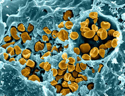
F. tularensis is a small Gram-negative bacillus, bipolar staining, better visible after Giemsa staining. Virulent strains possess a capsule.
C – Culture:
This bacterium can not be grown on the usual media that should be enriched with cysteine or thiamine and red blood cells (human, rabbit or sheep) (Francis medium, plain agar + cysteine, glucose and blood rabbit) or based on egg yolk. Culturing is carried out at 37 ° C under aerobic and colonies appear in 2-4 days. The risk of contamination from the cultures are very large and require special precautions. In practice, the direct diagnosis is never done except in specialized laboratories.
D – Identification – Experimental pathogenicity:
Depending on biochemical characteristics, two biovars are described, F. tularensis biovar tularensis biovar andpalaearctica.
Various animals, particularly laboratory rodents, are susceptible to experimental infection. The mouse is sensitive and all inoculation routes may be used including dermal single friction shaved skin.
This technique is useful for epidemiological studies or research from dead animals.
E – Indirect diagnosis:
Search agglutinating antibody is the essential element of laboratory diagnosis. The antigen is a suspension of killed bacteria. Antibodies appear from the 10th day, reaching a maximum as in 1 to 2 months (1/1000 or more) and will persist for years. There is an antigenic relationship with Brucella and cross-reactions were observed during brucellosis. Any presence of agglutinating antibodies, even at a low rate, must be confirmed by examining a second serum.
Search of hypersensitivity to tularine (autolysate heated bacteria) after intradermal injection (tularino feedback) is specific but is no longer feasible due to lack of reagent.
VI – TREATMENT AND PREVENTION:
Strains are sensitive to different antibiotics of the aminoglycoside, tetracycline, chloramphenicol, which are used for treatment.
Prophylaxis is based on the information of those exposed (hunters, gamekeepers, poachers!, Agricultural workers, …) especially when handling dead animals in infected area. It also relies on the health monitoring of imported hares Central European countries to the repopulation of hunting areas. Effective protection of exposed subjects is obtained with a vaccine based on live attenuated bacteria (used in the USSR).
You must be logged in to post a comment.