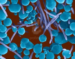
Cardiobacterium hominis
HISTORY:
Slotnick and Dougherthy proposed in 1964 the name C. hominis to designate gram-negative bacilli, polymorphic slow culture, initially placed in the group II-D (related bacteria Pasteurella) responsible exclusively endocarditis.There is no andgénique kinship with Brucella, Streptobacillus, Pasteurella and Haenwphilus.
I – HABITAT AND EPIDEMIOLOGY:
C. hominis is part of the normal flora of the upper respiratory tract and is present in the nose and throat. It is rarely found in the vagina. ‘
II – NATURAL PATHOGENICITY:
This bacterial species is exclusively responsible endocarditis.
C. hominis is a low virulent species. Endocarditis occurs when bacteremia point of departure and oropharyngeal transplant on pre-existing lesions.
III – BACTERIOLOGICAL DIAGNOSIS:
The levy is only represented by the blood culture is the only means of diagnosis of C. hominis endocarditis Positive culture is usually detectable after 1-7 days of incubation at 37 ° C (mean 4 days).; incubation should be extended 2 to 3 weeks.
Culture is manifested by a discrete flaky deposit without changing the clarity of the liquid medium.
IV – DIRECT EXAMINATION – MORPHOLOGY:
C. hominis is a right bacillus with rounded ends, sometimes swollen, 0.5-0.7 x 1-3 microns polymorphic, with long forms of 10-20 microns; bacteria are isolated, in pairs or in short chains grouped rosette packet pins.
It is a Gram-negative bacillus (with the particularity to retain the dye in its middle or blossom end), motionless, not encapsulated, non-spore forming.
V – CULTURE – IDENTITY CHARACTERS:
C. hominis is part of the optional aero-anaerobic group; certain strains require CO2 (3 to 5% CO2) in isolation.
Aerobically, strains need moisture. Growth is obtained 30-37 ° C.
The media should contain hemin, blood agar (rabbit, sheep, horse), chocolate agar; there is no culture on plain medium. Within 48 hours, the colonies are small, 1-2 mm, smooth, circular, convex, edged, opaque, non-haemolytic.
Bacteria have a fermentative metabolism acidifying various sugars (glucose, fructose, sucrose …) without gas production.
The main biochemical reactions oxidase (+), catalase (-), no reduction of nitrate to nitrite, ODC (-), urea (-) indole production in small quantities sought after extraction with xylene. The elements of the differential diagnosis are presented in the table.
VI – TREATMENT AND PREVENTION:
C. hominis is susceptible to antibiotics commonly used in combination for the treatment of endocarditis. Prophylaxis is that of bacterial endocarditis, in particular good oral hygiene.
Capnocytophaga

The genus Capnocytophaga was established in 1979 and brings together originally designated bacteria such asBacteroides ochraceus and DF1 group.
Three species, C. ochracea, C. gingivalis and C. sputigenatypically belong to this genre: they are fusiform bacilli Gram negative, cultivating anaerobic or aerobic conditions in the presence of 5-10% CO2 (hence the name: cell that eats carbon dioxide), fermenting glucose, giving pigmented yellow-orange colonies which have the particularity to spread and glide over the surface of the agar.
The taxonomic position of this kind has not been definitively established. It is presented here because of its habitat and its similarities isolated circumstances. C. canimorsus, previously designated as DF-2, has been linked to the genus Capnocytophaga. C. cynodegmi refers to strains that are close.
I – HABITAT AND EPIDEMIOLOGY:
These bacteria are part of the oral flora and were isolated at the gingival surface of the periodontal pocket, pharynx, saliva.
C. canimorsus is a commensal of the oral cavity in dogs and cats.
Human infection generally follows a bite.
II – PATHOGENICITY:
Capnocytophaga recognition as a pathogen for humans is recent. It has been isolated under particular circumstances.
A – dental pathology:
Although normally present at the mouth of the periodontal pocket of healthy subjects, Capnocytophaga seems responsible for certain forms of periodontitis.
B – Systemic Infections:
They are observed in patients with hematologic disease with granulocytopenia. They have also been observed in subjects without modification defenses.
C – Other Infections:
Isolated or accompanying bacteria oropharyngeal flora, Capnocytophaga is responsible for various infections: empyema, sinusitis, conjunctivitis, osteomyelitis …
III – PATHOPHYSIOLOGY. FACTOR VIRULENCE:
In immunocompromised patients, oral lesions or gengivales (gingivitis, ulcers), still present during sepsis, are the starting point of infection. In other cases, the simultaneous presence of bacteria in the oropharyngeal flora certifies the origin of the infection Capnocytophaga.
Differences in virulence between species have been observed in experimental studies of the periodontitis in animals.
The in vitro observed phenomenon of slippage at the surface of the medium may play an in vivo role in the infection of the periodontal pocket. More soluble bacterial substance alters the activity of leukocytes.
IV – BACTERIOLOGICAL DIAGNOSIS:
A – pathological products:
The specimens are varied and samples taken depending on the location of the infection; blood culture remains the most important collection.
B – Direct examination:
Bacteria of the genus are Capnocytophaga bacilli purposes elongated (0.4-0.6 x 2.5-6 um), fusiform, sometimes with a tapered end and the other rounded. These Gram-negative bacilli are not flogged, non-spore forming, not capped.
C – cropping Characters and identification:
Capnocytophaga is a facultative anaerobic kind that grows anaerobically, aerobically in the presence of 5-10% CO2, but not in ordinary atmosphere. The temperature should be between 30 and 37 ° C. The culture media are enriched media, blood agar (not essential), chocolate agar or media containing sugars. Some strains were isolated on Thayer Martin medium.
The colonies have a particular appearance due to spreading by sliding on the surface of the agar medium. This phenomenon of gliding motility, which leads Capnocytophaga to surround and cause colonies already on the environment is clean on blood agar sheep anaerobically; it is optimum with a medium in 3% agar.
Colonies are flat, 2-3 mm in 48 hours, the edges are irregular and spread in several directions. On blood agar, colonies are pink or yellow hue; the bacterial mass is yellow after sampling by scraping.
Some strains have colonies digging agar, others who adhere to the agar medium.
In liquid medium, nutrient broth containing sugars, culture results in a homogeneous turbid medium and fine grains culture or film adhering to the glass surface.
Biochemical characters oxidase (-), catalase (-), glucose fermentation and sucrose test ONPG (+), indole (-), LDC (-), ODC (-). Species are differentiated by sugar fermentation and nitrate reduction.
V – TREATMENT AND PREVENTION:
The species of the genus Capnocytophaga are usually sensitive to beta-lactam antibiotics (especially penicillins), tetracyclines, chloramphenicol, macrolides, lincosamides, quinolones and rifampicin; aminoglycosides, vancomycin, colistin and metronidazole are inactive.
Eikenella corrodens
E. corrodens, only species Eikenella corresponds to small gram-negative bacilli, slow and difficult culture, favored by hemin and CO2 fermenting glucose. This species, HB-1 group of King and Tatum, optional aero-anaerobicBacteroides corrodens is separated, became B. strict anaerobic ureolyticus.
I – HABITAT AND EPIDEMIOLOGY:
E. corrodens is present at the mucosal surfaces, mouth, upper airways, in dental plaque and in the intestine.
II – PATHOGENICITY:
A better understanding of this species has allowed its isolation in many pathogenic situations alone or associated with other bacterial species.
A – localized infections:
E. corrodens is responsible for the formation of abscesses in relation to contamination from the mucous membranes. Among the main locations should be mentioned:
– Abscesses of the brain and subdural empyema in sinusitis and dental abscesses in combination with a streptococcus;
– The pleuropulmonary infection with different bacteria;
– Dental infections periodontitis type;
– Skin infections after bite, or at hand when punched in the mouth, teeth and wound contamination by bacteria of the oral cavity, the infection is localized to the skin and subcutaneous tissue , to the joint or to the bone.
B – Bacteremia and endocarditis:
E. corrodens is responsible for endocarditis. After tooth extraction, bacteraemia are common but usually without clinical manifestations or consequences.
III – Pathophysiology – FACTORS VIRULENCE:
E. corrodens is mostly responsible for slowly progressive infection, indolent. The various maps are related contamination from the mucosa after injury or trauma. Its virulence is low, no toxin production has been demonstrated and if in some cases (endocarditis, meningitis, osteomyelitis) the bacterium is isolated only in the majority of cases of abscesses and suppurations E. corrodens is associated with another bacterial species (Streptococcus) and both species act in synergy.
IV – BACTERIOLOGICAL DIAGNOSIS:
A – pathological products:
E. corrodens can be isolated from many pathological Products: abscess, wound, joint fluid, blood culture, as well as sputum, nasopharyngeal swab, buccal, gingival. The cultivation is carried out on blood agar or chocolate that can be made selective by the addition of clindamycin.
B – Microscopic examination:
The bacterium is a small bacillus (0.2-0.5 x 1 -4 (Jm) Gram-negative coccobacillary at regular edges and rounded ends, not encapsulated, non-spore forming, immobile, sometimes forming filaments.
C – Culture – Identification characters:
Optional aero-anaerobic, E. corrodens needs Hemin (25 mg / 1) under aerobic conditions and a content of 5-10% CO2 promotes growth. The culture is carried out on blood agar or chocolate agar at 37 ° C; colonies cause greening of these environments. The moisture is favorable for cultivation. After 18 hours of incubation, the colonies are very small (0.5 mm) and will be more visible (1 mm) after further incubation. It is necessary to carefully examine the surface environments when a fast-growing species is present. Using a selective medium (clindamycin: 5 mg / 1) facilitates isolation from bronchopulmonary original samples.
A characteristic appearance (shared with other species such as A. actinomycetemcomitans, Capnocytophaga, C. hominis) is erosion, the excavation of the surface of the agar. This phenomenon detected by observing oblique light is not constant and the same strain may have variants without erosion (translucent colonies, smooth, curved);erosion is sometimes more visible by removing the settlements.
After 48 hours, the typical colony of E. corrodens on blood agar has three zones: a bright clear central area, a beaded circular area, speckled, highly refractive as multiple droplets of mercury, a non-refractive peripheral zone of active growth.
Depending on the conditions of observation, the colonies can also look like a bull’s-eye or a bug embedded in agar, or not to be confused with views as areas of condensation liquid desiccant (sometimes appearance largest coloniesCampylobacter).
These characters are less clear agar chocolate and vary depending on the incubation conditions.
The colonies, although that appear gray or translucent, have a more visible yellow pigment after prolonged incubation or involving several colonies.
In liquid medium, glucose broth, thioglycolate, culture E. corrodens manifested by adhering granular or flaky formations to the walls of the tube more or less reinforced in microaerophilic area. This feature is also common withA. actinomycetemcomitans and H. aphrophilus.
The main characters are identification oxidase (+), catalase (-), indole (-), reduction of nitrate to nitrite, LDC (+), ODC (+), urease (-).
V – TREATMENT AND PREVENTION:
E. corrodens is sensitive to many antibiotics: penicillins and cephalosporins (third generation), chloramphenicol, tetracycline, rifampicin, but it is resistant or insensitive to the first generation cephalosporins, methicillin, aminoglycosides, lincomycin (and clindamycin ) and metronidazole.
Aggregatibacter actinomycetemcomitans
This species was described by Klinger in 1912 under the name of Bacterium actinomycetemcomitans after its isolation in actinomycosis lesions (hence its name: accompanying an actinomycete). It is part of the genusActinobacillus alongside species pathogenic to animals.
I – HABITAT AND EPIDEMIOLOGY:
A. actinomycetemcomitans is part of the normal oral flora and is present at the plaque; infection by this bacterium are endogenous infections, usually oral starting point.
II – NATURAL PATHOGENICITY:
A. actinomycetemcomitans was isolated from lesions of actinomycosis associated with Actinomyces israelii andseems able to maintain the persistence of such lesions in the absence of Actinomyces Actinomycosis, but this role is not entirely clear.
A. actinomycetemcomitans is responsible for soft tissue infections sometimes suggestive of actinomycosis, endocarditis, abscesses. This bacterium is also involved in oral pathology, especially in periodontitis in adults and children.
III – BACTERIOLOGICAL DIAGNOSIS:
A – pathological products:
Blood culture is the essential consideration at endocarditis. Culture is detectable from the 3rd day of incubation and manifests itself in the form of granules accumulating bottom of the vial, the surface of the sedimented blood and adhering to the wall of the bottle.
Other levies are a function of location (brain abscess, soft tissue abscesses, cheek, face with A. israelii, otitis,sinusitis, bronchial pneumonia, tooth abscess), but isolation can be difficult because of the associated flora .
B – Direct examination – morphology:
A. actinomycetemcomitans is a small Gram-negative bacterium, immobile, non-spore-forming, non-encapsulated, or coccobacillary coccoid form. Bacillary forms can be observed after several subcultures. The bacteria are isolated, in pairs or contiguous rarely in chains. Colors are uneven.
C – Culture – Identification characters:
Culturing is carried out at 35-37 ° C in a CO2-enriched atmosphere. There is no requirement in X and V; the culture media were blood agar and chocolate agar.
On agar after 24 hours of incubation, the colonies are small with a diameter of 0.5 mm, but up to 2-3 mm in 5-7 days.The colonies are smooth, slightly domed translucent, with a wrinkled surface. After several days of incubation the colonies looks opaque center and have the appearance of a star with 4 or 6 branches leaving their mark on the agar when the colony is removed.
The colonies are adherent to the surface of the medium and are difficult to collect and separate.
In liquid medium, glucose broth, thioglycolate, the bacteria form granules which adhere to the surface of the tube, the remaining clear liquid. These colonies are difficult to collect and separate.
The main characters are biochemical oxidase (-) (except some strains), catalase (+), reduction of nitrate to nitrite, ODC (-), urea (-) (other species Actinobacillus possess urease) indole ( -), Fermentation of glucose but not lactose and saccharose.
D – Classification:
Pulverer and Ko described eight biotypes based on the fermentation of xylose, mannitol and galactose.
E – Other species Actinobacillus:
A. lignieresii, A. equuli, A. suis, A. capsulatus are species found in animals, normally present in the respiratory system, gastrointestinal tract or genital and leaders of various diseases in various animal species .
F – Treatment:
A. actinomycetemcomitans is sensitive to ampicillin, tetracycline, chloramphenicol …
Calymmatobacterium granulomatis
This very difficult growing bacteria is the only species of the genus Calymmatobacterium.
It was demonstrated in 1905 by Donovan as inclusion in mononuclear cells present in genital ulcers or “granuloma inguinale” (described in India in 1882).
I – EPIDEMIOLOGY AND PATHOGENICITY:
C. granulomatis is a bacterial pathogen only for man. Its habitat is poorly known and the epidemiology of the disease is poorly understood. It is responsible for “granuloma inguinale,” “Donovanosis” or “genital ulcerative granuloma.”This is a condition observed in countries with hot and humid climate on topics with colored skin. The locations are similar to those of chancroid. The lesions begin as a papule or nodule that evolve into chronic ulcers, indurated, granulomatous tissue formed hypertrophic velvety in the genital mucosa, without lymphadenopathy. The ulcers persist for months and the lesions spread via the lymphatics to the inguinal region. There are extragenital localizations of this condition, which for some, is not only a sexually transmitted disease.
II – PATHOPHYSIOLOGY:
Knowledge about the pathophysiology of this bacteria and virulence factors are very limited. There is a morphologically and antigenically related Klebsiella.
III – BACTERIOLOGICAL DIAGNOSIS:
A – The specimens:
The sampling is carried out at the ulceration in granulomatous tissue biopsy and fingerprints.
B – Direct examination:
The bacterium is a bacillus motionless, polymorph, capsule, Gram-negative, bipolar staining. Microscopic examination is the only element of diagnosis. Fingerprints and tissue smears are stained by Giemsa or Wright’s stain; bacilli is poorly other dyes and is not acid-fast.
Bacteria are isolated or in clusters in the cytoplasm of large mononuclear cells, constituting the “body Donovan”.Usually polymorphs, certain bacteria have a characteristic appearance hairpin closed security (due to the bipolar staining).
C – Culture:
The common areas and the usual enriched media do not allow the culture that has been obtained on medium containing egg yolk.
There are no other bacterial components as those gathered at the microscopic examination and the diagnosis of the disease is based on clinical appearance of lesions.
IV – TREATMENT AND PREVENTION:
The bacterium is sensitive to tetracycline, THIOPHENICOL®, erythromycin, co-trimoxazole, antibiotics that are used for treatment.


You must be logged in to post a comment.