HISTORY:
II there has almost 100 years of shaped bacteria turns as spirochetes were observed in the stomach of man or animals. There are about 50 years is known as the gastric mucosa has a urease activity capable of converting urea into ammonia. These observations are now explained by the existence of Helicobacter pylori.
The discovery of a curved bacterium that is a new bacterial species identified as Campylobacter and Helicobacter pylori seems to change many aspects of gastroenterology.
In 1979, Warren, an Australian author, observed on samples of antral mucosa curved bacteria were then called Campylobacter-like organisms. These bacteria were present in nearly all samples received and most of whom had histological appearance of chronic active gastritis.

Marshall and Warren constataient in 1982 that these bacteria are usually present in patients in which an indication of gastroscopy was raised and this presence was independent of the nature of the disease or symptom in question. The same authors then made a prospective study of 100 patients undergoing gastroscopy and biopsy of the mucosa. A curved Gram-negative microaerophilic and catalase positive was found in 95% of patients with chronic active gastritis. Isolates were positive in 70% of patients with gastric ulcer and 90% of patients with duodenal ulcer. For cons, the bacterium has been isolated from samples of healthy mucosa.
I – DEFINITION AND CLASSIFICATION:
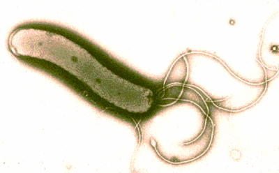
Helicobacter are gram-negative bacilli curved or C-shaped, U or S, by moving the presence April-June polar flagella.Bacilli are microaerophilic, difficult and oxidase growth (+).
Two species are described:
– H. pylori isolated from gastric mucosa of primates,
– H. mustelae, isolated from the gastric mucosa of the ferret, and he will not talk about below.
II – HABITAT AND EPIDEMIOLOGY:
Humans are the only known reservoir of H. pylori. The mode of transmission is unknown. H. pylori was found worldwide. It is insulated with particular frequency in the stomach ulcer or gastritis patients, even asymptomatic.
III – PATHOPHYSIOLOGY:
H. pylori is movable in the mucus. With adhesins it is capable of adhering to the cells of the lining of the stomach where it is found in the intercellular spaces.
Urease H. has pylori allows the production of ammonium ions can neutralize stomach acid, allowing the bacteria to multiply.
A cytotoxic protein produced by the bacterium prevent the goblet cells produce mucus protecting the lining of the acidity.
IV – PATHOGENICITY:
Several studies show that H. pylori plays an important etiological role in the type B gastritis (chronic active antral gastritis) and in the pathogenesis of peptic ulcer). The eradication of H. pylori to the mucosa is correlated with a decrease in the frequency of relapse of duodenal ulcer.
V – ISOLATION AND IDENTIFICATION OF H. Pylori
A – Sample:
Gastric biospsies are performed during a gastroscopy. Samples for bacteriological examination are either placed in saline at 4 ° C and seeded in time or placed in a suitable transport medium.
Benzocaine and simethicone were H. pylori inhibitory effect was not lidocaine.
The bacteria is not found in gastric fluid.
B – Identification of urease activity:
It can be searched directly or gastroscopy room or in the laboratory from a biopsy specimen.
C – Microscopic examination:
A homogenate of biopsy specimen is stained by the Gram method. Of gram-negative bacilli spiral or curved whose distribution is heterogeneous on the smear found.
A monoclonal antibody has also been used for the detection of H. pylori by direct immunofluorescence.
D – Culture:
Chocolate agar enriched horse blood Isovitalex 1%, incubated at 37 ° C in microaerophilic (10% CO2) and humid atmosphere of small (1 mm) transparent, smooth and even grow in 3 to 7 days.
The medium can be made selective by addition of vancomycin (6 mg / 1) and cefsulodin (5 mg / 1).
The identification is based on the morphology of bacilli Gram active urease, oxidase reactions and catalase positive.Alkaline phosphatase and GGT may also be highlighted. Other characters are negative.
VI – INDIRECT DIAGNOSIS:
The urease test (or breath test) is to make the patient to ingest the labeled urea ^ C and detecting after 20 minutes in the air expired labeled CO2 from the degradation of urea by the urease.
The detection of serum antibodies can be made by complement fixation, or immunoblot sensitized latex particles. But the interpretation is difficult since almost 50% of subjects 50 years of age have antibodies against H. pylori.
VII – TREATMENT:
The indications for treatment are still poorly defined.
H. pylori is sensitive to beta-lactams and aminoglycosides. It is resistant to trimethoprim-sulfamethoxazole. Bismuth subcitrate has a lytic action on H. pylori but is unable alone to eradicate the bacteria.
The recommended treatment combines ampicillin, metronidazole and bismuth subcitrate. Its effectiveness should be monitored by checking the eradication of the bacterium one month after stopping treatment.

You must be logged in to post a comment.