HISTORY:
The history of the discovery of this new bacterial genus is a model of epidemiological and bacteriological research by outstanding organization of the survey conducted in the field and the means deployed.
From 21 to 24 July 1976 was held at the Hotel Bellevue-Stratford in Philadelphia, the 58th congress of the American Legion (veterans).
Of the 4400 participants, 182 people (legionaries and companions) were suffering from lung disease, 15.9% died.Sixteen others presented findings of asymptomatic post. Identical cases (called “pneumonia of the high street”) were observed in people who were not entered in the hotel, but were found in the street near during the same month of July; Similarly, other cases were observed among participants in other meetings in the same hotel. The notion of an epidemic did not appear immediately because most cases only declared after the end of the congress and it was not until three days later that the practitioners of the city alerted health services, thus triggering extensive epidemiological investigation.
It then appeared that the contamination was done by air in the hotel lobby or on the sidewalk. The causative agent, infectious or not, was then actively sought. They looked so long a metal or other toxic agent in the tissue of deceased individuals; secondly, we employed 14 types of bacteriological or mycological media and 13 types of cell cultures in order to demonstrate a possible infectious agent.
Finally, on 18 January 1977, Mac Dade et al. announced a Gram negative bacterium was isolated from lung fragments from four of deceased subjects. These lung broyais were inoculated intraperitoneally to guinea pigs who presented a few days a fatal febrile illness. Liver smear exams and spleen showed numerous bacilli and inoculation of a homogenate of these organs in the yolk sac membrane of embryonated eggs dragged their death in 4 to 6 days.The etiological role of this bacterium in the epidemic in Philadelphia was demonstrated by indirect immunofluorescence on convalescent sera of the disease.
Subsequently, retrospective serological surveys allowed to attach to this disease several outbreaks of unexplained acute pneumonia.
In 1974 in Philadelphia, twenty people attended a conference at the Hotel Bellevue-Stratford eleven developed severe pneunopathie, two were dying.
In 1973 in Benidorm in Spain, eight Scottish tourists contracted lung infections, three died. They all stayed in the same hotel in July.
In 1965 in Washington DC, there were in the psychiatric hospital St. Elizabeth 81 patients with severe pneumonia which 14 were dying. The infection had touched topics sleeping with the window open on the park and those walking in the same park where earthworks were carried out.
In 1968 in Pontiac, Michigan, there were 144 cases of benign acute febrile illnesses among employees and customers of a health center. Infection was synchronized with the starting of the air conditioning system.
In 1943 Tatiock had isolated a strain of L. micdadei as the “Rickettsia-like.”
So we found that he was not a new disease. Finally, in 1980, this bacterium was named Legionella pneumophila.
I – TAXONOMY – CLASSIFICATION:
Legionella bacteria belong to a new family: the Legionellaceae.
The generally accepted classification is that of Brenner family Legionellaceae currently has only one kind:Legionella, with nearly thirty species identified by DNA / DNA hybridization study and partition chromatography gas liquid (Table I). The type species is L. pneumophila is the most common medical condition and has 14 serogroups.
Some authors mention clear differences among the species of Legionella bacteria and talk about neighboring referred Fluoribacter (bozemanae F., F. dumoffii. F. gormanii) Tatiockia (L. micdadei, L. maceachernii).

II – ECOLOGY:
A – Habitat:
Legionella are aquatic bacteria mundane of our natural environment. They are found in water (preferably hot) where survival is long: lakes, rivers, marshes, wetlands, water pipes; in air: water vapor, aerosols, nebulizers.
The study of legionellosis cases has shown that the presence of the organism is related to the
near earthworks of water tanks, generated aerosols
or by the cooling towers of air conditioning systems, or by
shower heads, taps the large open …
These bacteria nevertheless demanding cultivation conditions may grow, reaching concentrations in the order of 106 bacteria per liter. It seems that the presence of a microscopic flora associated (Pseudomonas, Flavobacterium,protozoa …) facilitates the growth of Legionella. It particularly emphasizes the role of free-living amoebae(Acanthamoeba and Naegleria genera) which, by ingesting without destroying Legionella, seem to provide the perfect conditions for survival and dissemination.
These bacteria are killed by heat above 65 ° C and the chlorination of water.
B – Epidemiology:
It has been shown that the Legionella (mainly L. pneumophila serogroup 4) were present in 60% of samples of water carried in town.
Moreover, the prevalence of Legionella pneumonia is modest (about 4%), so it seems that the concept of land is very important.
The presence of Legionella in a water sampling should therefore not worry, except in immunocompromised Service (grafted blood diseases) where water sterilization should be considered.
D Note that there is no transmission from one individual to another and the healthy carrier is exceptional.
To monitor human infections, legionellosis is now a reportable disease (see Appendix).
III – PATHOGENICITY:
A – pathogenic natural power:
Human infections are usually due to L. pneumophila (90% of cases), other species are rarely involved and their pathogenicity still poorly understood.
Legionella generally occur in a particular field. It affects mostly men over 50, smokers, ethyl, persons infected with chronic cardiopulmonary disease, chronic kidney disease or diabetes, subjects in a state of cellular immunosuppression .
1. usual clinical form: Legionnaires’ disease
Incubation lasts 2 to 10 days. After a gradual onset, the patient has the status phase intense infectious syndrome and respiratory disease (pneumonia extensive poorly systematized), often associated with pleural effusion.
In 1/4 of the cases, there is no focal neurological signs (headache, confusion, seizures). Lumbar puncture is usually normal. There is rarely meningitis, encephalitis, polyradiculoneuropathy. The most frequently advanced mechanism is that of a neurotropic toxin.
Digestive disorders are common (especially diarrhea).
In the biological picture, there are often laboratory evidence of liver and kidney damage; there is also hyponatraemia and hypophosphatemia.
The diagnosis of legionellosis is to raise with a very broad febrile pneumonia associated with extrapulmonary manifestations (diarrhea) occurring in a privileged field for which the usual bacteriological examinations are negative.
2. Other clinical forms:
a / Legionella immunocompromised (nosocomial legionellosis)
Transplant subjects, patients with leukemia trichorhinophalangeal leukocytes, lung cancer or are at very high risk groups. For subjects with AIDS, genital or breast cancer, the risk seems lower.
The etiology usually plurimicrobienne and fragile terrain that changes are often very serious with a very high fatality rate (about 40%).
b / Pontiac fever:
It is a benign affect the upper respiratory tract caused by a particular strain of L. pneumophila serogroup 1.
The incubation period is short (36 hours), the chest x-ray are normal healing occurs spontaneously in a few days.
c / subclinical forms are frequent:
Serological studies show that over 10% of the population has a high rate of AAU-pneumophila serogroup 1 antibodies.
d / Other forms:
hematologic, renal, brain, skin, heart (artificial valve) or muscle.
B – Experimental pathogenicity:
Guinea pigs inoculated intraperitoneally develops a febrile illness progressing to death within days.
Is observed during the first 4 days peritonitis, then the seed passes through the blood vessels and lymphatics.Necrosis homes then appear in the spleen, liver, lungs where we find these organisms in large numbers.
Bacilli are phagocytosed by monocytes-macrophages, cells in which the Legionella can survive. Lysis of these immune cells is the source of the microbial release.
A delayed-type hypersensitivity (DTH) reaction has been demonstrated in guinea pigs sensitized after inoculation of live bacteria or killed by heat.
The HSR is detected by the protein antigen common to various serogroups of L. pneumophila.
This bacterium has an endotoxin which the in vivo activity is much lower than that of other Gram-negative bacteria. L.pneumophila, L. micdadei, L. bozemanii possess a virulence antigen mip (macrophage infectivity potentiator), the gene carried by a plasmid has been cloned and sequenced.
IV – LABORATORY DIAGNOSIS OF LEGIONELLA INFECTIONS:
A – Withdrawals used and transport conditions:
The search can be performed on bronchoalveolar lavages or preferably on transtracheal aspirates, lung biopsies, pleural fluid centrifugation the pellets, blood cultures, fragments of various organs, possibly urine, feces.
All these samples will be collected as sterile as possible to avoid contamination. To prevent desiccation be added a little sterile water (do not use saline which may inhibit the culture). Similarly, during the sampling, it is best to avoid local anesthetic (lidocaine).
If the laboratory in transport time exceeds 30 minutes, it is advisable to keep the sample at 4 ° C. If the delivery is delayed more than 24 or 48 hours, the sample will be frozen at -20 ° C minimum. These precautions are valid for crops and for searches by direct immunofluorescence.
B – Legionella Research by direct immunofluorescence in pathological Products:
1. Principle:
Using antibodies directed against various different serogroups and species of Legionella. Û is also a monoclonal antibody specific for the species Legionella pneumophila which recognizes a protein antigen located on the outer membrane of all known serogroups of L. pneumophila. These antibodies are conjugated to fluorescein.
In a patient treated for less than 3 days, the review is still possible though less sensitive, but reading is more difficult, the bacterial body lose their normal morphology. In addition there is a background fluorescence due to bacterial lysis.
The IFD can be performed on fresh or preserved in formalin or in paraffin.
2. Practical realization:
Several smears is carried out in a thin layer, one was stained by Gram technique appécier the dominant flora. Note that the Legionella are not displayed by the usual Gram staining. This bacterium is Gram stained using carbol fuchsin basic. It then appears Gram negative.
For the IFD is generally used 3 pools fluorescent conjugates:
– A: L. pneumophila serogroups 1, 2, 3 and 4.
– B: L. pneumophila 5 and 6, L. dumoffii, L. longbeachae 1.
– C: L. gormanii, L. micdadei, L. longbeachae 2, L. bozemanii.
The review will be said to be positive if one sees little more than 5 frankly fluorescent bacilli (apple green) and having a typical morphology (often curved, polymorphs). These bacilli are intra- and extra-leukocyte.
3. Interest – Limits:
The major advantage of this technique is its speed (about 1 hour). Its sensitivity is poor and function of the examined sample (30-50%).
A negative review should therefore not be out this diagnosis, it is mandatory to grow crops.
Specificity is good, but there may be some cross-reactivity with Pseudomonas, Streptococcus pneumoniae, Escherichia coli, Bacteroidesfragilis. The specificity is enhanced by the use of monoclonal antibodies.
C – Culture and isolation:
1. Culture media:
Legionella can be grown on the usual media. Various media were used successively. Currently is no longer used as Buffered Charcoal Yeast Extract supplemented with alpha-keto glutaric acid or alpha BCYE incubated at 35 ° C under 2.5 to 5% of CO.
This medium is made selective by the addition of various antibiotics. Usually a mixture vancomycin, cefamandole, polymyxin B, anisomycin.
For environmental sampling was recommended to add a mixture: glycine, vancomycin, polymyxin (GVP medium).
In preparing these environments, it is essential to pay particular attention to the quality of the ingredients and their addition order and the final pH conditions that must be strictly between 6.85 and 6.95.
The guinea pig inoculation has long been used. Its results are delayed.
2. Practical realization:
Before sowing, it may be useful to decontaminate the multi-microbial samples such as tracheal aspirates or bronchoalveolar lavage. To do this, the sample is treated with HC1 buffer – KC1 at pH 2.2. In the case of infection byPseudomonas aeruginosa, it will treat the sample by the heat (1 to 2 minutes at a temperature less than or equal to 60 ° C).
Each sample is then seeded on 6 medium boxes BCYE-alpha: 3 Single BCYE-alpha and alpha 3 selective BCYE.Each of the boxes of the two media is inoculated with the pure sample, diluted to 10-2 and 10 “4 in distilled water to reduce the concentration of natural inhibitors present in the products, antibiotics and possible contaminants.
Regarding blood samples, Legionella can grow in special environments for blood culture (Bactec).

3. Isolation and identification:
a / Morphology and structure of bacteria:
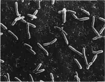
They are short bacilli or Cocco bacilli January-February microns long and 0.5 to 0.7 pm wide, strictly aerobic.
In vitro, filamentous bacteria is observed (up to 100 microns long).
They are movable by a single polar flagellum.
In vivo, they may be intracellular.
b / Characters crop:
It is a difficult bacillus culture growing slowly (2-15 days on average, and even higher in blood cultures). The growth is optimum at a temperature of 35 ° C, the limits between 25 and 48 ° C.
It is a strict aerobic germ, demanding cystine and iron, which grows better in the presence of 2.5 to 5% CO.
The Petri dishes are examined daily (for 15 days) with a dissecting microscope. When colonies appeared, they said, a typical aspect fried glass and can be pigmented (from blue to pink depending on species), pigment more intense UV light. This typical aspect, but nonspecific is generally observed that for 24 h. All these colonies are picked and replated on BCYE alpha devoid of cysteine and on blood agar.
An identification of Legionella can then be made when the following arguments are met:
– Typical aspect of culture BCYE-alpha,
– Absence of culture on BCYE alpha-L-cysteine-free, except for L. jordanis and L. oakridgensis.
– Absence of culture on blood agar.
The species identification may sometimes be performed using different biochemical characteristics. We note the species still positive character (catalase) and always negative characters: acidification of carbohydrates, nitratase, urease (Table II). Confirmation will often require the analysis of branched fatty acids by gas chromatography (GLC), ubiquinones HPLC or the study of the genome by DNA / DNA hybridization.

c / Interest isolation:
Only the isolation of the germ provides absolute diagnostic certainty. In addition, the isolation of Legionella is the only diagnostic biological argument when the IFD and serology were negative.
The comparison of strains isolated from patients and their environment has a major epidemiological interest to determine the exact origin of the contamination.
D – serology by indirect immunofluorescence (IIF):
1. Principle:
The antigen is either Legionella grown on embryonated eggs and killed by formalin (Taylor antigen, the most widely used in Europe due to its greater sensitivity) or Legionella grown on artificial media and killed by formalin and heat ( Wilkinson antigen).
The antigen is fixed by acetone slide. The technique used is that of the usual IFA using a total human anti-gamma-globulin conjugated fluorescent.
2. Practical realization:
Is removed first as soon as possible serum and a serum every 8 days (for 6 to 8 weeks sometimes) to observe a possible sometimes late seroconversion.
Taking into account the number of species and serogroups of Legionella, it is physically impossible to test in routine vis-à-vis serum antigens corresponding to all the known species of Legionella. This serological study is facilitated by the existence of cross-reactions between different serogroups of L. pneumophila and cross-reactions between different species of Legionella.
In practice, use of pools of antigens L. pneumophila 1-4 and 5-6 as well as the monovalent antigen L. 1pneumophila. If vis-à-vis a positive antigen pool, a titration is carried out vis-a-vis each monospecific antigens.
For. pneumophila and using the Taylor antigen, the diagnosis is considered certain if the antibody level rises of less than 1/16 to more than 1/64 th and very likely when the title is stable at 17128 e.
3. Interest:
He often used to confirm a clinical suspicion of diagnosis and is sometimes the only way to confirm a diagnosis.
The sensitivity is around 80%. With Taylor’s antigen specificity is good. Note that false positives are (Chlamydia andMycoplasma, Pseudomonas).
Search IgM has no interest in this status.
E – Other techniques:
1. Detection of antibodies agglutination slide:
This easy to use technique is beneficial for L. pneumophila with results close to the IFIs; but there are many cross-reactions between other Legionella species.
2. Detection of soluble antigens by ELISA:
Soluble antigens of L. pneumophila 1 can be detected in serum and urine using ELISA using polyclonal and monoclonal antibodies.
The sensitivity in the urine is close to 80% and specificity of about 99%.
3. Other detection methods:
The in vitro gene amplification could soon lead to improved direct diagnosis of this bacterium.
V – TREATMENT – ANTIBIOTIC SENSITIVITY:
In vitro, L. pneumophila is sensitive to rifampicin, cefotaxime, erythromycin, aminoglycosides, tetracyclines, chloramphenicol, penicillins, new quinolones. This germ often produces an active beta-lactamase on cephalosporins.
These data do not coincide with the clinical results and the classical treatment is based on erythromycin intravenously currently associated with rifampicin. Fluoroquinolones are increasingly used alone or in combination.
VI – CONCLUSION:
Legionella currently account for 5-10% of the etiologies of atypical pneumonia, and as such are responsible for an adverse outcome in a number of immunocompromised patients.
Their isolation is now within the reach of every good laboratory that wants to empower. By cons, isolation is long and tedious, it is necessary that these review requests are justified by a real clinical suspicion.
You must be logged in to post a comment.