HISTORY:
Staphylococci were found in pus by Pasteur in 1880. In 1883, Ogston created the “Staph” to describe these grains (Kokkos) grouped in irregular clusters in the manner of a bunch of grapes (Staphylos). In 1884, Rosenbach obtained pure cultures of these bacteria.
He divided the genus Staphylococcus into two groups depending on whether the colonies were white or brown.
1 – GENERAL CHARACTERISTICS OF STAPHYLOCOCCI:
A – Taxonomic Position and Classification:

The Micrococcaceae family is composed of three kinds of Grani positive cocci in clusters that differ in their G + C%:Staphylococcus (30-39%), Micrococcus (65 – 75%) andPlanococcus (48-52%). This genre is met only marine bacteriology.
The species in these three genera possess catalase and grow aerobically. The Gram-positive cocci in clusters that grow only under anaerobic conditions are referredPeptococcus and will be treated with anaerobic bacteria.
Genus Staphylococcus occupies a very important place in human and animal pathology. The genus Micrococcus a pathogenicity virtually zero.
However micrococci strains are frequently isolated in medical bacteriology. It is then of contaminants as distinguished from staphylococci. The characters that distinguish the Staphylococcus and Micrococcus are shown in Table I.

– () Species or strains with a different character
a: S. hominis is strictly aerobic and S. strict anaerobic saccharotyticus
b; Micrococcus kristinae and Mr. varions can grow anaerobically
c: M. kristinae Mr. varions can ferment glucose
d: S. sciuri, S. lentus, S. caseolyticus have cytochrome c oxidase
Within the Staphylococcus genus are distinguished according to the classification of Kloos and Schleifer more than 20 species. A number of them are found in humans, others are present in animals or in foods (meat, dairy products …). Among the species found in humans:
– Three species occupy a privileged place: 5. aureus, S. epidermidis and
S. saprophyticus
– Other, more rarely involved in human disease are: S. hominis
S. haemolyticus, S. warneri, S. capitis, S. saccharolyticus, S. auricularis,
S. simulate, S.cohnii, S.xylosus, S. intermedius, S. and S lugdunensis. schleiferi.
It is conventional to oppose S. aureus produces coagulase and is often pathogenic staphylococci other, non-producing coagulase and rarely cause infections.

B – Habitat and epidemiology:
It is very common germs in nature (air, water, soil).Staphylococci, in particular the species S. aureus andS. epidermidis, are part of the normal flora of many individuals who are “asymptomatic”. However, these strains may be the source of infection or auto-contaminate other individuals.
It is estimated that 20-75% of individuals are carriers of S. aureus persistent carriers, holders occasional or temporary; in contrast, some individuals are “non-carriers”.
Staphylococci can be found especially in the anterior nares (S. aureus: 30 – 40%, S. epidermidis 30-100%).They can also be isolated from the skin (S. epidermidis 85-100%) and especially hot, humid areas it (axilla, perineum) where you can find S.aureus. n is not uncommon to isolate S. aureus stools.
Transmission is mainly direct human to human (contact, hand-borne spread from the nose in particular) or indirectly through food or surroundings.
The newborn was quickly colonized by S. aureus after childbirth and, later, the child can be contaminated within a community; the person transmission is very variable, some individuals being “dangerous disseminators” while other subjects almost never transmit their strains.
II – STAPHYLOCOCCUS AUREUS:
A – Pathogenicity:
1. Natural pathogenic power:
Infections with S. aureus are frequent and appear very different clinical aspects.
a / The cutaneous staphylococcal infections, subcutaneous and mucous membranes:
– S. aureus may cause superficial or deep skin infections.
The superficial infection results impetigo, folliculitis or a onyxis.
Deep infection is represented by intrafollicular abscesses all the hair sheath called furuncles, or by infection of the channels of sweat glands called hidrosadénites. Anthrax is a conglomeration of boils and malignant staphylococcal infection of the face is a particularly serious localization.
Furunculosis and hidradenitis may be recurrent. Recurrences are sometimes associated with triggers diabetes, overwork …
There is also skin infections related to the presence of catheters as well as psoriasis or eczema superinfected by S.aureus, but without clinical signs of infection.
– The rashes scarlatiniform part of toxic shock syndrome associated with the presence of the toxin-producing strains of toxic shock syndrome or certain enterotoxins. Some rashes are also linked to the presence of pyrogenic toxin.
– Lyell’s syndrome or staphylococcal disease Ritter von Rittershain or scalded child syndrome is related to the secretion of a staphylococcal toxin, or exfoliatin épidermolysine that causes flaking of the surface layer of the epidemic.
– At the mucous membranes, S. aureus may be involved in phlegmons amygdala, sinusitis or recurrent otitis sometimes.
b / Locations visceral S. aureus:
They occur either in isolation or in the context of overt sepsis:
– Bone staphylococcal infections: acute osteomyelitis is a condition of the child or young person; it typically affects the long bones and can become chronic.
Post-surgical bone staphylococcal infections are very worrying.
– Staphylococcia pleuropulmonary: infant forms are very frequent and severe. The adult forms are rare and can appear after a viral infection such as the flu.
– Staphylococcia Urogenital: staphylococcal pyelonephritis are fairly common; S. aureus can also cause isolated abscess formation kidney or perirenal phlegmons.
– Staphylococcia neuromeningeal: they are rare and dominated by meningitis observed especially in neuro-surgical environment (valves) S.. epidermidis is then often isolated. Meningitis should not be confused with epidural staph, pus then being located in the epidural space.
Brain abscess may be encountered.
– Staphylococcal endocarditis occurs particularly in patients with artificial heart valves. Among drug addicts, they are often right heart endocarditis.
c / Septicemia S. aureus:
The staphylococcémies are caused and maintained by a complicated primary infection site thrombophlebitis; are frequent infections, a worrying gravity. Evolution is enamelled septic metastases.

d / Foodborne infections:
– They are due to the ingestion of enterotoxins (A and E), preformed in the food, resistant to the digestive juices and some heat, causing early onset disorders (less than 3 hours) with vomiting, diarrhea, dehydration and absence of fever.
– The evolution is benign, except in cases of significant fluid and electrolyte losses (elderly, infants).
– Staphylococcus must be sought in the food.
– The search for enterotoxin in foods or produced by the isolated strain is possible in specialized laboratories.
and acute enterocolitis:
– They occur during the course of antibiotic therapy and are due to the proliferation of S. aureus enterotoxin AROs and producer.
– These staphylococci should be sought in the stool.
– Specialized laboratories can detect whether the isolated Staphylococcus enterotoxin producers or not.
// Toxic Shock Syndrome (TSS)
It combines fever, hypotension, rash erythematous macular followed by desquamation scarlatiniform often diarrhea.Blood cultures were sterile.
It is linked to the action of staphylococcal toxin TSST-1 (Toxic Shock Syndrome Toxin-1), or, it seems, certain serotypes enterotoxin.
This syndrome was described in 1978 in the ILS.A. as the result of a vaginal proliferation S. aureus in women using tampons. Officials strains of TSS can also be isolated from various injuries.
The high incidence of TSS in the USA between 1978 and 1982 has not been observed in Europe.
2. Experimental pathogenicity:
It is necessary to inject 5.106 CFU of S. aureus under the skin to produce healthy skin infection in humans; by 100 against bacteria enough on a suture zone or with pre-existing skin lesions.
No laboratory animal is not able to reproduce different aspects of the human disease. But the rabbit is the most sensitive animal: subcutaneous injection of S. aureus produces an abscess that spontaneously heals and intravenous injection leads to death in 4 to 10 days with visceral abscesses (especially kidneys).
B – Pathophysiology:
S. aureus can become pathogenic as a result of various circumstances:
– Penetration of the germ in the body, most often after breaking the skin barrier (injuries, surgery, burns, dermatitis, injections, catheters, …) or at a hair follicle.
– Disruption of the host bacterium balance following circumstances favoring infection: viral infection (influenza, measles), antibiotic selecting a strain of S. aureus, immune deficiency, diabetes, alcoholism …
It follows a bacterial multiplication with production of enzymes and toxins corresponding to the expression of the virulence of the organism. This explains the spread of infection that can lead to sepsis. Schematically, several steps can succeed where are involved various original substances staphylococcal:
– Local invasion: hyaluronidase exfoliatin,
– Cell necrosis: proteases, lipases, esterases, DNase, phosphatase, alpha toxin (and beta, gamma, delta)
– Reduction in local defenses: Leukocidin, capsule, protein A,
– Regional thrombophlebitis home: coagulase
– Septic emboli: plasmin.
Because of its many toxins and enzymes, S. aureus destroys the cells of the body and produce pus. It is thus the same type of pyogenic germ.
C – Substances excreted by S. aureus:
They are particularly numerous in S. aureus.
Genetic determinants are known for many of these substances (Table II).
1. S. aureus toxins:
– Hemolysins: their main properties are summarized in Table III:
– Alpha-hemolysin or staphylococcal alpha-toxin protein in nature, heat-stable antigen, inducing the formation of neutralizing antibodies.
It is cytotoxic and cytolytic for a wide variety of cell types.
It appears to be inserted into the cytoplasmic membrane of target cells and permits passage of small molecules.
– Beta-hemolysin is thermolabile. It acts as a type C sphingomyelinase and gives increased hemolysis in the presence of Streptococcus agalactiae: the CAMP test (named after their discoverers: Christie, Atkins, Munsch-Petersen)
– Gamma-hemolysin has two factors 1 and II acting synergistically.


These are antigenic and cholesterol inhibits their action. The molecular mechanism of action of this toxin is unknown.
– Delta-haemolysin contains hydrophobic and hydrophilic amino acids and acts as a detergent to the membranes.
Panton Valentine leukocidin destroyed very specifically granulocytes and macrophages in human and rabbit. The purified leukocidin causes a dermonécrose rabbits. The toxic effect on leukocytes is due to a modification of the cation permeability. There are 2 F and S components acting synergistically.
– Enterotoxins: they are responsible for food poisoning and are characterized by their molecular weight between 27,800 and 34,100 Daltons, their isoelectric point and serotyping.
These number of 7: A, B, D and E.
Serotypes A, B and D are the most common in food poisoning.
Some of these enterotoxins have a mitogenic effect on T cells
Some (Ent B) are more thermostable than other proteins. They are resistant to proteolytic enzymes.
The minimum toxic dose is around 1 (g per 100 g of food. The detection of toxins in food, feces or broths is by an immunological technique ‘(immunodiffusion, agglutination, radio immunology, ELISA).
The possession of enterotoxin gene is not exceptional. Include one or more of these toxins in about half of hospital strains, which makes difficult the connection of a clinical syndrome of isolation of enterotoxigenic strain.
– The épidermolytiques toxins (or exfoliatines) are produced by certain strains of S. aureus (5%).
These are proteins with a molecular weight of 27 000 daltons and antigenic; 30-40% of phage group II strains produce these toxins, or 5% of hospital strains.
One distinguishes two types A and B: the gene encoding the serotype A is chromosome (90% of exfoliatines) and that encoding the serotype B is plasmid (4-5% of exfoliatines). The 2 serotypes may be produced by the same strain.
These toxins (A or B) cause intraepidermal cleavage. The mode of action at the molecular level is unknown. These toxins can be detected on newborn mice by immunoelectrophoresis against or ELISA. Toxigenic strains are particularly responsible for neonatal and childhood infections: Lyell’s syndrome, bullous impetigo staph …
– Pyrogenic toxins. There are two pyrogenic toxins, mitogens, and nonspecific antigenic a molecular weight of 12,000 daltons divided into two types A and B. The pyrogenic effect is observed on the rabbits. These toxins are involved in staphylococcal scarlatiniform fevers.
– Toxin toxic shock syndrome (TSST). In this particular syndrome is observed erythematous rash with or without desquamation. Responsible staphylococci produce a toxin (TSST-1) sensitive to proteolytic enzymes, antigenic and a molecular weight of 20 000 daltons; this syndrome has also been associated with enterotoxin production; This toxin is produced by 90% of the strains isolated in toxic shock syndrome and 11% of hospital strains-run.
– “Succinic oxidase factor.” The toxin inhibits the succinate oxidation by isolated mouse liver mitochondria.
2. Enzymes of S. aureus:
These enzymes have a pathogenic interest and / or important diagnostic.
a / Coagulase free:
S. aureus is able to excrete a protein causing coagulation of human plasma or rabbit (derived citrate, oxalate, heparin or EDTA) and called “free coagulase.” Staphylococcus producing this toxin will be identified as S. aureusBut two species observed in veterinary practice can produce this enzyme. S. intermedius and S. hyicus.
The formation of the clot does not require the presence of calcium, it is not activated by the purified fibrinogen, but needs a neighboring globulin prothrombin (coagulase-reacting-factor). This is antigenic coagulase (7 antigenic groups) and causes the appearance of antibodies that inhibit the biological activity.
It plays a role in suppurative thrombophlebitis of the formation and inhibit phagocytosis.
In the laboratory, the detection of coagulase occurs by bringing the rabbit plasma and the strain to be studied in a tube at 37 ° C; caking of the mixture is carried out in 3 to 6 or sometimes 24 hours; coagulation may be followed by a dissolution of the clot as a result of the action of staphylokinase.
b / Coagulase bound or dumping factor or affinity factor for fibrinogen:
Alongside this coagulase “free”, we recognize an insoluble coagulase, “tied” to the surface germs and also called dumping factor. It binds to fibrinogen and is responsible for the aggregation slide staphylococci with serum; this affinity factor for fibrinogen can be demonstrated by contacting the strain to be studied with sheep red blood cells or latex particles coated with fibrinogen; agglutination in seconds is found in 98% of strains of S. aureus. The dumping factor does not degrade fibrinogen into fibrin. However, this test can be positive for some species of coagulase-negative staphylococci, including S. lugdunensis and S. schleiferi.
c / Fibrinolysin or staphylokinase:
This enzyme is a plasminogen activator (action similar to that of streptokinase), acting on the human and rabbit plasma. This activity is demonstrated on agar plates containing fibrin (clearing zone).
This thermolabile substance is antigenic. It dissolves clots and may play a role in septic emboli formation.
d / Hyaluronidase:
This thermolabile enzyme hydrolysis of hyaluronic acid, it fluidizes the ground substance tissue conjonctifet can be searched by viscometry.
e / Nuclease:
Nuclease (DNase) S.aureus (or thermonuclease) is thermostable, while that of other bacterial species is thermolabile.
Enzyme activity is highlighted on DNA based medium with toluidine blue (pink halo); The reaction can be made more specific by serum inhibition of nuclease S aureus, search can be performed in 4 hours.
// Other enzyme activities
Lipase activity, esterase, protease can be highlighted (medium egg Baird-Parker), and a phosphatase activity; and there are 2 phosphatases (acid and alkaline) it is easy to search.
D – Structure and antigens:
Several substances staphylococci wall play a role in immunological reactions.
1. The peptidoglycan:
Staphylococci and micrococci have peptidoglycan which differ in their amino acids. S. peptidoglycan structureaureus is well known: this diamino acid is L-lysine.
Staphylococci are lysed by an endopeptidase, lysostaphin, which cleaves glycyl-glycine bond present in the interpeptidique bridge, but they are generally resistant to the action of lysozyme muramidasique.
2. teichoic acids:
The wall of staphylococci comprises polyolphosphates (polyribitol or polyglycerol-phosphate) substituted hexose.

Micrococci contain teichoic acids without ribitol or glycerol.
S. teichoic acids aureus are also called pA polysaccharide. Under certain conditions the teichoic acids can be replaced by teichuroniques acids (N-acetyl-D fucosamine, N-acetyl-D mannosaminuronique acid polymers in S. aureus.) The teichoic acids play an important role on the interactions between bacteria and cells and attachment of bacteriophages; also they are antigenic. They are covalently linked to the peptidoglycan chains.
3. Surface Polysaccharides:
These surface antigens have been described in encapsulated strains. This capsule can be formed more abundantly during the growth of S. aureus in the presence of serum in a weakly agar medium. These antigens are also detectable in many strains say noncapsulated but carriers of identical polysaccharides in smaller quantities. Some of these polysaccharides prevent the activation of the alternative complement pathway and thus protect the bacterium from phagocytic and bactericidal activity of the serum. A classification of these polysaccharides 8 capsular types was proposed and two types 5 and 8 cover 70-80% of the strains responsible for septicemia; this work may lead to the detection of staphylococcal soluble antigens.

4. Protein A:
II is a protein (MW 42 kDa) antigenic, insoluble in the native state and constituting the wall of S. aureus. It sets (Figure 3) the Fc portion of human immunoglobulin IgG, 2 and 4, leaving the Fab free part. In vitro, and can easily attach to the surface of S. aureus immunoglobulins (eg anti-meningococcal or anti-pneumococcus.) for detecting the corresponding antigens in cultures or pathological material; it is coagglutination.
NB: fixing IgG on protein A is a function of the animal species from which the IgG and also come from the sub-classes IgG considered: for example, IgG sheep or goat bind bit on the protein TO.
5. Antigens type and serotyping:
All strains of S. aureus have surface antigenic factors used in classification. There are nearly 30 antigenic factors and two main classifications.
In Pillet system 13 sera are used; the most common serotypes I, II, III, 18; in France 10% of non-typeable strains in this system.
The Oeding system allows a better approach to the antigenic structure (mosaic) of strains, but is more difficult technical achievement; antigens are designated by the initials a4, a5, bl, cl, hl …
6. Phage typing:
This method is used to compare strains of S. aureus isolated during hospital outbreaks. It consists in subjecting the strain to be tested in a series of 23 bacteriophage. Lysis of the strain by a bacteriophage is conditioned by the existence of the wall at the level of a specific receptor for the phage. Some lytic combinations occur more frequently than others, which helped define phage groups: group I, II, III, IV (not observed in humans) and V.
Groups 1 and III are the most common in men.
E – Bacteriological diagnosis of S. aureus infection:
1. The levies:
They should be done before any antibiotic therapy and practiced with strict asepsis: not only for blood cultures, CSF, urine, but also for the pus, biopsies, bronchial aspirates, swabs. Avoid contamination of the disease produced by strains of Staphylococcus aureus and S. often epidermidis on the skin. Repetition allows blood cultures to rule in favor of sepsis or contamination.
Bacteriologists may also be required to seek and to enumerate staphylococci in food, water, or air in hospitals.
2. Direct examination:
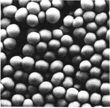
Staphylococci are Gram-positive cocci, 0.8 to 1 micron in diameter, arranged in clusters, in diplococci, short chains or clusters in typical. They are motionless, asporulés sometimes encapsulated.
Observation of Gram-positive cocci in short chains or clusters in pus often evokes a staphylococcus.
3. Cropping characters:
S. aureus grows abundantly on agar medium (colonies of 1 to 2 mm in diameter); some strains produce a yellow-orange pigment, but this production is irregular.
Culture is obtained in 18 to 24 hours at 37 ° C (crop possible from 10 to 45 ° C) on regular. S. media aureus grows in the presence of high salt concentrations (selective medium Chapman 7.5% NaCl). The optimum pH is 7.0 to 7.5.
There is demanding variants of growth factors: thiamine, pantothenic acid …
For monomicrobial product isolation is easy in broth or in solid non-selective medium (Trypticase Soy, Mueller-Hinton blood agar).
For pathological polymicrobial products or food, one must resort to selective media such as the middle of Chapman (hypersaline environment + mannitol) or middle of Baird-Parker tellurite (mainly used in food microbiology).
4. respiratory Type:
S. aureus is an aerobic-anaerobic germ optional.
Some strains require CO2 to grow. All strains produce catalase.
5. Identification:
In 18 to 24 hours on rich agar medium, colonies were observed 1 to 2 mm in diameter may produce a yellow pigment and consist of cocci Gram (+) in clusters, catalase (+).
On the middle of Chapman, S. aureus causes acidification (turn yellow) of mannitol; in the middle of Baird-Parker, 5.aureus forms black colonies (reduction of tellurite) with a clear halo (proteolysis) and, later, a clouding in the halo (lipase).
Suspicious colonies be sought classical coagulase or inhibition of thermostable nuclease using antibodies. Rapid screening tests also exist (research of the affinity factor for fibrinogen and protein research A). But there are some false positives and false negatives with these tests.
If it is of the species S. aureus identification is sometimes supplemented by an epidemiological perspective further galleries, by serotyping or phage typing.
To note:
– The isolation of some strains of S. aureus from blood cultures in particular, can be difficult (endocarditis, chronic suppurations) because some strains give slow onset microcolonies whose growth can be enhanced by the addition of growth factors.
– The search for toxins: enterotoxins exfoliatin is reserved to date to specialized laboratories but enterotoxin research by passive agglutination techniques of latex particles or ELISA, directly in the products are now used, including food bacteriology.
– The term pathogenic staphylococci (like that of Staphylococcus aureus) should be outlawed: S. aureus isolated from the skin or mucous membranes (carry) is not pathogenic, while S. epidermidis may, in certain circumstances, be pathogenic.
6. Differential diagnosis:
-. Micrococcus This germ is separated from staphylococcal his respiratory type, its inability to oxidize glycerol (1%) in the presence of erythromycin (0.4 (ig / ml) and by its resistance to 200 ug / ml lysostaphin (see Table I).
– Other species of staphylococci: it should be noted that some strains of S. aureus may not produce free coagulase due to mutation.
In other strains of S. aureus, the affinity factor for fibrinogen can be masked by the presence of a polysaccharide capsule.
Of staphylococcal species other than 5. aureus may be pigmented particularly in primary culture: S. saprophyticus, S. hominis, S. haemolyticus …
Other species may be hemolytic (S. haemolyticus, S. warneri, S. schleiferi …)
Finally species other than S. aureus can grow on medium Chapman and acidify mannitol (S. cohnii, S. saprophyticus, …).
-. Stomatococcus This germ, catalase negative or low, does not grow on salt medium, is not hemolytic and elastic consistency colonies adhere to the agar.
7. Rapid Diagnosis:
It does not currently exist rapid and reliable diagnosis of S. aureus search of soluble antigens. The best candidates for this type of antigen screening would be represented by surface polysaccharides of Staphylococcus.
8. Indirect diagnosis:
It currently has little practical value.
Titration of alpha antistaphylolysines may be of interest in deep or chronic infections (including bone), the standard rate is less than or equal to 2 IU per ml. The value of this serological reaction is being challenged.
The search for anti-teichoic acids or anti-peptidoglycan by immuno-electrophoresis or against gel diffusion may have indications if endocarditis or metastatic complications with negative blood cultures outbreak or inaccessible to culture, but does not have to date well standardized antigen.
III – STAPHYLOCOCCI coagulase negative:
A – Identification:
Coagulase negative staphylococci are identified completely when the circumstances of isolation indicate that they are potentially in a position to play a pathogenic role. IV and V tables show the bacteriological features that make the diagnosis of species.

About anaerobic growth, hemolysis and test novobiocin, three points should be made.
1. Study the growth of anaerobic (inoculation with 0.5 ml of broth Me Farland 0.5 units):
S. hominis hardly grows in aerobic (thioglycolate broth).
S. saccharolyticus only grows in anaerobic conditions.
S. Haemolyticus and S. capitis grow much better aerobically and anaerobically.
2. Study of the rate of appearance of hemolysis:
Hemolysis S. haemolyticus is late (after 24 hours of often oven).
3. Test novobiocin:
S. saprophyticus is resistant to this antibiotic. This test is useful on single urine strains. But other staph can also withstand novobiocin (S. cohnii, S. xylosus).
In practice, S. hominis, S. haemolyticus and S. simulate represent 5 to 20% isolated coagulase-negative staphylococci in humans.
B – Clinical significance of coagulase-negative staphylococci:
Some species (S. epidermidis, S. hominis, S. saccharolyticus, S. capitis and S. auricularis) are consistently found on the skin or mucous membranes natural orifices.
Other species are encountered inconstantly and density of these bacterial populations is often low. There is an ecological adaptation of S . capitis scalp and S. auricularis the external auditory meatus.


Coagulase negative staphylococci have long been considered as non-pathogenic and as mere contaminants defective samples. Today it is clear that at least two species, S.epidermidis and S. saprophyticus, are potentially pathogenic opportunistic bacteria.
Main infections caused by coagulase-negative staphylococci, and their place in pathology are shown in Tables VI and VII
1. S. epidermidis:
It may be responsible for vascular or prosthetic joint infections, heart valves, cerebrospinal fluid shunts valves. He is also involved in the occurrence of consecutive peritoneal dialysis peritonitis, subacute endocarditis in drug addicts, and a variety of endophthalmitis particularly in immunocompromised infections. The ability of this species to colonize the surface of the polymers (catheters, prosthetic devices), and the cells would be related to the abundant polysaccharide capsule produced by this organism.
2. S. saprophyticus:
This species has a name particularly inopportune because it may be responsible for urinary infections that have the particularity to observe in young women usually not hospitalized. This species is responsible for 5 to 10% of urinary tract infections due to its ability to adhere to urinary epithelium.
3. The other species:
They have a very casual pathogenicity or even questionable. To assert their role in infection, it is necessary to isolate the strain on several occasions in the absence of other potentially pathogenic bacterial species.
C – toxic factors developed by staphylococcus coagulase – negative:
These staph can develop such as delta hemolysin, and sometimes related coagulase a plasmin, a Leukocidin a DNase. Practically, strains isolated from infections (wounds, blood cultures, abscess) frequently produce one or more of these virulence factors that strains isolated from healthy subjects.
IV – TREATMENT AND PREVENTION OF INFECTIONS STAPHYLOCOCCI:
A – antibiotic treatment:
Antibiotics have modified the prognosis of serious infections such as malignant staphylococcal infection of the face.Systemic staphylococcal infections, whether S. aureus or S. epidermidis, should be treated with bactericidal antibiotics. The bactericidal activity is generally obtained by combining penicillin group M antibiotic to another family.The resistance of a strain to all beta-lactam justifies the use of antibiotics such as vancomycin or teicoplanin that are constantly active, but are also more slowly bactericidal and more expensive.
1. Beta-lactam problem:
a/ The secretion penicillinases:
– phenomenon Description
Today nearly 90% of staphylococcal strains isolated in hospital and non-hospital environment resistant to penicillin G production penicillinases that open the beta-lactam ring of the molecule and inactivate the antibiotic. These penicillinases are extracellular, inducible and usually encoded by plasmids.
They inactivate G and V penicillins, aminopenicillin the carboxypénicillines and ureidopenicillins.
By cons they have little affinity for methicillin, oxacillin, cloxacillin and all cephalosporins which remain active on these strains producing penicillinase. These penicillinases are inhibited by clavulanic acid.
– Detection of enzymatic resistance
The penicillinase production is detected by observing the standard zone of inhibition around a penicillin antibiogram disc G. It results in a reduction of inhibition of the diameter with respect to a sensitive strain not producing penicillinase. A sensitive strain has a large diameter of inhibition with a fuzzy edge, called ghost area, which corresponds to a lysis of the bacteria. A resistant strain by producing penicillinases gives a smaller area to clean edge inhibition. At the periphery of the zone of inhibition are well developed colonies called “squatter settlements”.
In practice, the penicillin G is sufficient disk on susceptibility testing to detect resistance to all penicillins. Strains having a penicillinase must be considered resistant to these antibiotics. We should not make an income sensitivity as “intermediate”.
Where doubt remains on the fact that a strain occurs or not penicillinase, detection can be made by testing Gots acidimetric test, or by a iodometric test or simply by the nitrocefin test.
b/ Intrinsic resistance or heterogeneous methicillin resistance:
– phenomenon Description
This non-enzymatic mechanism of resistance to beta-lactams has been observed since 1961, when the introduction of methicillin in therapy. Strains that possess the expressed heterogeneous methicillin resistant or “ori R”. In a ori R strain, only a small proportion of the bacteria is capable of expressing resistance and grow in the presence of methicillin.
The ori R strains should always be considered resistant to all beta-lactam antibiotics, including third-generation cephalosporins and imipenem.
It is also producing penicillinase. They are usually resistant to other antibiotics as aminoglycosides, tetracyclines and macrolides.
10 to 40% of hospital strains of S. aureus isolated in France ori R.
Their frequency among non-hospital original strains is low.
This resistance is a consequence of changes in enzymatic proteins involved in the synthesis of peptidoglycan cell wall. There is in S. aureus at least four PLP (Liaison Proteins Penicillin) also called PBP (Penicillin Binding Protein).Among the ori R strains there is a reduction in the affinity of PBP for beta-lactams and synthesis of an abnormal PBP PBP 2a whose affinity for beta-laetamines is low.
– Detection of heterogeneous resistance
There are homogeneous strains resistant to methicillin high. Detection by susceptibility poses no problem.
Resistance is often heterogeneous and only a small fraction of the bacteria is able to express resistance in culture standard conditions. To promote the expression of resistance to methicillin susceptibility on it is necessary to place a methicillin disc (or oxacillin which is more stable)
– On a Mueller-Hinton agar plate incubated between 25 and 30 ° C and observed after 24 and 48 hours.
– Either on an agar plate of Mueller Hinton hypersaline (5% NaCl) and incubated at 37 ° C.
Under these conditions, the heterogeneous resistance translates the presence of small colonies in the zone of inhibition around the disc. These colonies are more visible after incubation for 48 hours
A methicillin resistance has to be considered as a strain resistant to all beta-lactams. Clinical experiments have shown that these strains were indeed resistant to cephalosporins. In practice, the susceptibility it is useful to test a single disk: methicillin or oxacillin.
The heterogeneous resistance speaking evil around cephalosporin discs, there is no need to use them.
In case of doubt the highlight of the intrinsic resistance susceptibility it is interesting to use agar dilution technique. A dilution of the test strain (containing 104 CFLJ spot) is seeded on Mueller-Hinton agar plate in 5% NaCl containing 10 mg / 1 of methicillin or 6 mg / 1 oxacillin. The growth of the strain on the box after 24 to 48 h at 35 ° C is considered resistant to all beta-lactams.

c/ Tolerance:
Beta-lactam antibiotics are bactericidal antibiotics. The minimum inhibitory concentrations (MIC) and minimum bactericidal concentrations (MBC) are normally neighbors.
The report CMB / MIC of the order of 1 to 2.
Tolerance is a particular resistance mechanism in which the MIC is normal but where the strain is not lysed by highly above the MIC CMB. A tolerant strain is called when the WCD is at least 32 times higher than the MIC.
This phenomenon applies to all involved in antibiotic biosynthesis wall (beta-lactams, but also vancomycin, fosfomycin), with sometimes as cross tolerances between the beta-lactams and vancomycin. This tolerance is related to an intrinsic peptidoglycan hydrolase system inoperative (defective). Tolerance is phenotypic, that is to say, reversible and unstable. Strains of S. tolerant aureus are mostly found in endocarditis; the current frequency and the significance of such strains are not well established.
2. Other antibiotics:
a/ Aminoglycosides:
They can be modified by various staphylococcal enzymes: phosphotransferase, and nucleotidyltransferases acetyltransferases.
The resistant strains are also resistant to gentamicin, tobramycin and kanamycin (phenotype KTG) and have a reduced sensitivity to netilmicin and amikacin, irrespective of the diameters of inhibition observed for these two antibiotics. In addition, the WCD amikacin is much higher in these strains. In practice, the strains of S. aureusresistant to gentamicin should be considered resistant to all common aminoglycosides (except streptomycin and neomycin must be tested separately).
Strains resistant to tobramycin and kanamycin (phenotype KT) are also resistant to amikacin and neomycin, but remain sensitive to gentamicin and netilmicin.
Strains K Nm phenotype (resistance to kanamycin and neomycin) are also resistant to amikacin.
These strains resistant to aminoglycosides (particularly KTG phenotype) are usually ori A.
b/ Vancomycin and teicoplanin:
Constantly active on S. aureus same ori R, they are used only in hospitals, particularly in endocarditis and severe infections. The susceptibility of coagulase-negative staphylococci teicoplanin is less constant and must be verified in the laboratory.
c/ Macrolides:
In hospitals, about 25% of strains of S. aureus resistant to erythromycin and a percentage slightly lower resistance to lincosamides (lincomycin, clindamycin). Less than 5% of the strains were resistant to synergistins (pristinamycin, virginiamycin).
Resistance can be constitutive, that is to say, not induced, and concerns macrolides, lincosamides and streptogramin B (resistance Streptogramins A are not met and synergy between and persists in vitro sensitivity to pristinamycin and virginiamycin seems preserved.
Some strains of S. aureus exhibit resistance mlsg induced by erythromycin or oleandomycin. These strains are sensitive to some macrolides (spiramycin, josamycin, midecamycin), lincosamides and streptogramin A and B. They are resistant against erythromycin and oleandomycin.
The combination of traces of one of these two antibiotics to one of the other antibiotics of the MLS group induces an identical mlsg resistance constitutive type.
The isolated resistance phenotype alone lincomycin or associated with resistance to streptogramins A (LS ^) is still rarely seen in France.
d/ Chloramphenicol, cyclins and cotrimoxazole:
They can not be considered antistaphylococcal choice.
el Rifampicin:
She is very active but should always be used in combination to prevent the selection of resistant mutants.
// Other antibiotics
They just take an interesting place among antistaphylococcal antibiotics, even to treat the ori R strains, provided they are used in combination; these are:
– Fusidic acid
– Fosfomycin (95 to 100% of susceptible strains)
– Fluoroquinolones (pefloxacin, ofloxacin, ciprofloxacin …).
In practice, where the bacteriostatic antibiotic is sufficient (ENT infection, cellulitis, boils, …) can be used products pristinamycin type, but for serious infections (septicemia, endocarditis, pneumonia, osteomyelitis, toxic shock, …) is must obtain a bactericidal effect can be used:
– If the strain is ori S, a beta-lactam Association (oxacillin or cephalothin) – and aminoglycoside – if the strain is ori R, associations of vancomycin and aminoglycoside with or without rifampin, or pefloxacin associated with fosfomycin or with an aminoglycoside or cefotaxime-fosfomycin sometimes. The choice is then guided by studies of bactericidal activities of associations in vitro, and possibly by the study of the bactericidal power of the serum on the strain involved.
Strains belonging to species other than S. aureus are often multiresistant ori and R (35 to 66% of strains); they are all susceptible to vancomycin.
Note that fosfomycin is still inactive S. saprophyticus.
The largest therapeutic difficulties are encountered with endocarditis or meningitis S. epidermidis, and in these cases the study in vitro associations is required.
Finally, note that some strains of staphylococci can acquire vis-à-vis antiseptic resistance especially mercury, which can promote the spread of such strains in hospitals.
B – Prophylaxis:
1. Individual:
The healthy carrier does not constitute a danger to the subject itself. But in a chronic furunculosis carrier subject, it is necessary to eliminate this port.
In a scalable staphylococcal lesion, avoid sepsis. Vaccine trials based staphylococcus toxoid or have not led to convincing results.
2. Collective:
It should theoretically prevent the use of germ carriers in catering.
The fight against hospital staph is a fundamental problem of hospital hygiene based on the observation of aseptic procedures, staff education and rationalization of the use of antibiotics.
You must be logged in to post a comment.