HISTORY:
The genus Yersinia, named after the bacteriologist Alexandre Yersin who first isolated in 1894 the plague bacillus, was proposed in 1944 and formalized in 1974 after the breakup of the genre Pastewella.
In 1883 Malassez and Vignal isolated Pasteurella pseudotuberculosis in guinea pigs with pseudo-tuberculous lesions of the liver, spleen, and lymph nodes; this bacterium was responsible for animal diseases especially in rodents and the role of Y. pseudotuberculosis in human pathology was shown in 1954.
In the 1960s, Y. enterocolitica was responsible for animal diseases reaching chinchilla farms in Western Europe and has since then been isolated in a growing number of human cases. Currently, the plague bacillus no longer occupies the place that was his for centuries and the center stage is occupied so much less dramatic V.enterocolitica.
I – GENERAL CHARACTERISTICS OF GENRE:
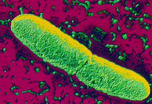
 The genus Yersinia includes bacilli rights, sometimes cocco-bacilli, Gram-negative, with a tendency to bipolar staining, 0.5-0.8 (-a x 1-3 um, not encapsulated, non-spore forming, immobile 37, moving below 30 ° C by a ciliature peritrichous. However Y. pestis is still immobile.
Yersinia present the general characteristics ofEnterobacteriaceae which the genus though with features such as the expression of phenotypic traits temperature dependent.
II – TAXONOMY AND NOMENCLATURE:
The genus Yersinia is composed of several species that can be separated into two groups: one species virulent Y.pestis (Yersin bacillus), Y. pseudotuberculosis (Bacillus Malassez and Vignal) and Y. enterocolitica (serogroup 0: 3 0: 8 0: 9 and 0: 5.7); and secondly the non-virulent species for humans: Y. enterocolitica of other serogroups, Y.intermedia, Y. frederiksenii, kristensenii Y., Y. aldovae. Y. ruckeri pathogenic to fish, do not belong to the genusYersinia. Two new species, Y. mollaretii and Y. bercovieri have recently been described. Y. pestis and Y.pseudotuberculosis are two closely related species genetically (very high homology hybridization DNA-DNA) and could be two pathogenic variants of the same species.
III – HABITAT AND EPIDEMIOLOGY:
Bacteria of the Yersinia genus have a wide distribution and can be isolated in the soil and in many animal species.Pathogenic species reach various animal species and occasionally humans.
A – The plague bacillus:
Y. pestis is present mainly in rodents. Dozens of species present on all continents can be infected. The spread of the disease is provided by the rodent-chip torque and epidemiology of plague is closely related to the ecology of rodents and their fleas. The bacteria survive for months in soil, and contaminated by dead bodies burrows of rodents and fleas are the bacteria tank. When the animal disease reached rodents cities, especially rats, contamination of man becomes possible through the flea bite. The plague disappeared from Europe at the beginning of the century but is still observed in Africa (Central, East, South), Asia (South East, USSR, Iran) and in North America and South, in enzootic form.
Y. strains pestis have particular biochemical characteristics depending on their geographical distribution: biovarantiqua is observed in Central Asia and Central Africa; the medievalis biovar met in Iran and the USSR; the biovarorientalis (or oceanic) has a worldwide distribution.
B – Y. pseudotuberculosis:
This species as a reservoir soil and contaminated animals from the ground like rodents and birds. Other animal species may be involved. The disease in humans is observed during the cold season after direct contact with carrier animals that eliminate Y. pseudotuberculosis in feces. Animals close to man as a cat (rodents hunter and birds) and small rodents “leisure” kept at home play a significant role. The disease is also observed in the spring after contamination from the soil or contaminated food or plants by the feces of mammals or birds. This makes the infection by Y. pseudotuberculosis a pseudotellurique saprozoonose, common disease in animals and humans with a common reservoir formed by the external environment.
C – there enterocolitica.:
This species has a distribution and a much larger tank. Moreover heterogeneity which will be described biochemically also exists epidemiological perspective. Y. enterocolitica was isolated from rodents, small mammals, pigs, in water, soil, food. There are strains adapted to an environment or a host (human or animal) and non-adapted strains found in soil, water, the digestive tract of mammals.
Y. enterocolitica is pathogenic to chinchillas, rabbits, monkeys and humans.
The biovar 5 strains are encountered in the hare in Europe, biovar 3 in the chinchilla. The strains of biovar 4, serogroup 0: 3, biovar 3 serogroup 0: 5 and biovar 2 serogroup 0: 9 are the most common in men in Europe; those of serogroup 0: 8 are more frequently isolated in the USA
Human disease occurs mostly in 1960 and the increase in the prevalence of infections with Y. enterocolitica in humans can be attributed firstly to the peculiarity of the species to multiply at low temperature and the other to the frequent presence of Y. enter ocolitica in some foods (vegetables, meat and sausage, milk and derivatives). This is related to the growing importance of the cold chain for food preservation within the family, with changes in eating attitudes and consumption of many raw vegetables, with the spread of catering that amplifies both foregoing.
Therefore contamination is mainly through the digestive tract; exceptionally, subcutaneous, ocular, and after cat scratches were observed.
Y. species intermedia, Y. kristensenii and Y. frederiksenii present in the environment (water, soil) and occasionally isolated from humans are considered non-pathogenic.
IV – PATHOGENICITY NATURAL:
A – Y. pestis:
It is the agent of plague, a disease primarily wild rodents animal animal transmitted by the chip. In the latter the bacteria multiply and engorge the esophagus and pharynx; contamination occurs during the meal at which the chip regurgitates the bacteria.
Man is an accidental host chosen by Xenopsylla cheopis (rat flea) when no other available host (where rat populations are decimated). Other arthropod can rarely serve as vectors.
Infection after flea bite results in the typical bubonic form. The bacteria multiply locally and is spread via the lymphatic system. The nodes of the territory of the bite, inflamed and enlarged, is the bubo. The disease progresses in septicemic sometimes with secondary pulmonary localization.
The human transmission can be by dispersion from bronchopulmonary secretions of a subject with secondary pulmonary localization. She then drives in the new subject and contaminated primary pulmonary form. This mode of infection by aerosol could exceptionally be observed when working in the laboratory.
B – Y. enterocolitica and Y. pseudotuberculosis:
They are responsible for yersiniosis, bringing together various pathological manifestations. Infections Y.enterocolitica are the most common.
1. Digestive Events:
It is the oldest known and far the most frequent (older children, adolescents and adults). The mesenteric adénolymphite is the most classic form observed with both species, reflected in a painful acute abdomen in the right iliac fossa, which may be confused with acute appendicitis. The infection as acute ileitis is less common.
In young children (up to 6 years), Y. enterocolitica is responsible for gastroenteritis whose clinical picture is similar to that observed with other intestinal bacteria include fever, abdominal pain, diarrhea.
2. Septicemia:
The septicemic forms are rarer and are observed in a particular field (cirrhosis, diabetes, hemochromatosis, blood diseases, …). Systemic infections are more common in subjects with iron overload. The usual clinical sepsis may be accompanied by secondary locations (liver, stomach, lymph node).
Because of its ability to grow in globular concentrated pockets stored at 4 ° C Y. enterocolitica may be responsible for transfusion consecutive septic shock bacteremia in the donor.
3. extra-digestive Events:
They are caused most often by an autoimmune process:
– Erythema nodosum occurring after infection Y. pseudotuberculosis in children between 8 and 15 years or after infection with Y. enterocolitica in women after age 50,
– Arthritis and reactive arthritis succeeding one episode of acute enteritis but for the most part carriers of HLA-B27 antigen.
Other events have occasionally been described based on serological arguments syndrome Fiessinger Reiter, carditis, glomerulonephritis, thyroiditis.
Fever scarlatiniform Far East due to Y. pseudotuberculosis and described for thirty years in the easternmost part of the Soviet Union has not yet been observed in Europe.
V – Pathophysiology – FACTORS VIRULENCE:
Pathogenic Yersinia have in common several virulence factors.
Y. enterocolitica was the first species to which the invasive capacity was related to the presence of a plasmid, now common mechanism Shigella, Salmonella and entero-invasive E. coli strains. al.
Virulence plasmid (pYV) of 70 kb is present in pathogenic strains.
Several plasmid genes encode the outer membrane proteins (Yop), the V antigen or W; the synthesis of these proteins is carried out at 37 ° C in the absence of calcium ions, but no or little at 28 ° C. These proteins help to resist phagocytosis.
PI protein produced independently of calcium ions is made up of subunits covering in the form of fibrils, the surface of the bacteria. Encoded by pYV, this protein would support the adhesion and protect the bacteria from the bactericidal action of the serum.
Two chromosomal genes responsible for the invasion were identified. One, cat, present in Y. pseudotuberculosisand Y. enterocolitica encodes a protein of 103 kDa or “invasin”, produced at 28 ° C on the surface of the bacteria and in the outer membrane. Another chromosomal gene of invasion is the garlic gene (locus of attachment and invasion) that encodes a 17 kDa protein produced at 37 ° C. Only pathogenic strains possess garlic while the inv gene is present but not functional in non-pathogenic strains (environmental) Y. enterocolitica.
The highly virulent strains, Y. pestis, Y. pseudotuberculosis, Y. enterocolitica 0: 8, possess high molecular weight proteins (190 kDa) synthesized in an iron deficient medium. The gene is absent in other species and in Y.enterocolitica 0: 3 and 0: 9.
Y. enter ocolitica product at 28 ° C enterotoxin with identical properties to those of the heat-stable enterotoxin (ST) ofE. coli. The role of enterotoxin in the pathogenesis of the infection is unclear because it is not produced at a temperature above 30 ° C.
The urease by mucosal changes produced by ammonia released from the intestinal contents, participate in bacterial colonization.
Many factors will thus participate in the virulence of Y. enterocolitica and Y. pseudotuberculosis. The achievement of the lamina propria would be done through specialized epithelial cells without brush border overlying the Peyer’s patches (M cells) or through the enterocytes.
In laboratory animal species are virulent Y. pestis, Y. pseudotuberculosis and Y. enterocolitica (serotype 0: 8 0: 9 and 0: 3).
In humans, especially in children, infection with Y. enterocolitica carries a picture of gastroenteritis and signs of systemic infection. Bacteria causing diarrhea as a enteroinvasive mechanism. The front door is located in the terminal ileum and caecum at which the main lesions are observed in the form of inflamed areas and ulceration in the region of the Peyer’s patches. The bacteria are intracellular multiplication in the cells of the mucous forming inflammatory granulomas and micro-abscesses that progress to necrosis causing ulceration and bleeding.Mesenteric lymph nodes are also the seat of bacterial multiplication in mononuclear cells with inflammatory granuloma formation; mesenteric adenitis can sometimes present a pseudotumoral appearance.
VI – BACTERIOLOGICAL DIAGNOSIS:
A – The specimens:
The special case of the plague will not be considered here. The main samples are feces, mesenteric lymph nodes (especially for Y. pseudotuberculosis), appendix, blood for blood culture and other sampling according to location.The samples can be kept cold, which, for the products plurimicrobiens is used as enrichment medium.
B – Direct examination – Body type:
Yersinia are small Gram-negative bacilli, sometimes coccoid whose morphology is similar to that of Pasteurella. A bipolar staining is detectable, it is more pronounced for Y. pestis and Y. . pseudotuberculosis Bacteria are movable to 22-29 ° C but still at 37 ° C Y. pestis is still immobile.
C – Culture – cropping characters:
Culture is possible on ordinary media (nutrient agar). In 24 hours at 37 ° C, colonies are most often at the limit of visibility; they increase in size by extending the incubation and can be identified much more easily. All species, after 48 hours of incubation, exhibit some polymorphism of the colonies.
Y. enterocolitica and Y. pseudotuberculosis are psychrophilic species and can grow at temperatures between + 4 and + 10 ° C.
Uncontaminated samples are cultured on conventional media. Research Yersinia in plurimicrobiens products (faeces, food, environment …) enrichment methods may be used, especially incubation at low temperature (4 ° C) for several days or weeks in liquid medium (water peptone, PBS).
Looking for Y. enterocolitica by stool is facilitated by the use of selection media. Selective media containing bile salts (MacConkey agar, Hektoen, Wauters, SS, DCL) are used and incubated at 30 ° C. There are also selective media made by the addition of antibiotics (CIN medium: cefsulodin, irgasan, novobiocin) for detecting low amounts of Y.enterocolitica in stool.
Y. enterocolitica is present in the stool in diarrhea and can persist for several weeks or months after clinical recovery.It can be searched remotely acute events, especially during autoimmune attacks. Against by Y. pseudotuberculosisdoes not persist in the gut after the cessation of the diarrhea episode and is no longer found in the stool occurs when mesenteric adenitis.
D – Identification:
Yersinia present the general characteristics of Enterobacteriaceae: they are Gram-negative bacilli, optional aero-anaerobic fermenting glucose oxidase negative and reducing nitrate to nitrite (except Y. pestis var medievalis and biovar 5 Y. . enterocolitica).
Important elements of identification are: the lack of mobility at 37 ° C and mobility below 29 ° C. Depending on the strain, mobility can not be observed in isolation and can not appear after several subcultures.
The reaction of the urease is still substantially and positive (except Y. pestis).
The test ONPG is positive but the strains do not possess beta-galactosidase.
The absence of lysine decarboxylase, arginine dihydrolase, of phénylalaninedésaminase is constant. The production of H ^ S and culture on Simmons citrate are negative.
It should be stressed thermo-dependent appearance of phenotypic especially for Y. enterocolitica. Some characters such as the production of acetoin or acid will be more clearly and quickly positive after incubation at a temperature below 30 ° C. The principal characteristics of the species of genus Yersinia are presented in Table I.
The biochemical heterogeneity of Y. enterocolitica can define five chemotypes (or biovars) on the basis of variable characters (indole production, fermentation of xylose, the presence of a lipase, reducing esculin) (Table II).
There is not enough sensitive animal species that can assist in the identification of strains of Y. pseudotuberculosisand Y. enterocolitica. The main elements of differential diagnosis before a negative lactose colony, and positive for urease based on the search for specific characters to Proteus (PDA or ADD, production of H2S). Other species such as Enterobacter will differentiate Y. enterocolitica. Finally the other species of Yersinia, there is little regarded as atypical strains of Y. enterocolitica are sometimes less easily identified (see Table I).


E – Classification – serotypes – phage types:
The antigenic structure of Yersinia is complex; they have common antigen Enterobacteriaceae. Some antigens are virulence determinants (fraction 1 envelope antigen, V and W antigens) and have been discussed in the relevant chapter.
The strains of both species, Y. pseudotuberculosis and Y. enterocolitica serotypes are classified according to their specific somatic antigens.
Y. pseudotuberculosis has been divided into six types (I to VI) according antigens 0. chemotype 1 is the most common. There are cross-reactions between type II and group B Salmonella, type IV and group D Salmonella, type VI and E. coli 055. There are 5 flagellar antigens (a to e).
In Y. enterocolitica antigens 34 0 20 H antigens have been described. They define serogroups useful for epidemiological studies. A small number of serogroups is associated with a human or animal disease. A change in the distribution of types of strains isolated in humans stool is observed since 1985. Indeed, if 0: 3 is still the most common, 0: 9 seems in decline and there is a significant increase in stem chemotype of 1 (0: 6; 0: 5 …), deemed non-pathogenic in authentic diarrheal syndromes.
There is a particular geographic distribution; Stem 0: 3 and 0: 9 are found in Europe, whereas strains 0: 8 are the most common in the US.
There are cross-reactions between Brucella and Y. enterocolitica 0: 9.
Epidemiological studies can be completed by phage typing and there are two classification schemes, one of which, French, recognizes 10 phage types (I to X).
F – Indirect diagnosis:
Serological research is useful for diagnosis in extra-intestinal manifestations. They explore the presence of agglutinating antibodies or complement fixing antibodies. The antigens used are the type of antigens 1 to V for Y.pseudotuberculosis and antigens 0: 3, 0: 9 0: 5 Y. enterocolitica in an agglutination reaction tube or microagglutination. ELISA is used.
Greater than or equal to 200 titles by conventional agglutination technique and 40 by microscopic agglutination are considered significant.
Cross reactions between type 0: 9 and Brucella are observed with both methods. Other antigenic cross-reactions especially with Salmonella group B or group D, and the presence of antibodies in apparently healthy subjects are the difficulty of interpreting the routine serology. It should in all cases to track specific antibodies. The antibodies increase a week after the onset of symptoms and reach their peak in the second week of illness.
VII – TREATMENT AND PREVENTION:
Y. pseudotuberculosis is usually sensitive to antibiotics active against Gram-negative bacilli: beta-lactams, aminoglycosides, tetracyclines, Y. enterocolitica is naturally resistant to ampicillin and first generation cephalosporins by producing both a constitutive beta-lactamase and beta-lactamase (cephalosporinase) inducible.Rare strains have acquired resistance to other antibiotics. As for the biochemical characteristics, the expression of the resistance is dependent on temperature.
In severe infections, antibiotics will be chosen from those mentioned above or among the third generation cephalosporins associated or not with an aminoglycoside.
There is no specific prevention of yersiniosis.
You must be logged in to post a comment.