A- esophageal squamous cancer: definition, incidence, epidemiological and etiological factors
Squamous esophageal cancer, like other cancers of the upper aerodigestive sphere develops from the squamous epithelium.
Its incidence is relatively high in France, particularly in the western regions (incidence of 30 per 100,000 inhabitants), where the primary role of alcohol and tobacco has been shown, which explains its male.
In areas of very high incidence (Asia, South Africa) where the deficiency factors appear to be involved, the distribution is the same gender.
We know the other disease states esophagus predisposes to this type of cancer caustic esophagitis, achalasia cardia.
B- Adenocarcinoma of the esophagus: definition, incidence, epidemiological and etiological factors
Its prevalence is about 20 times smaller than the squamous cell carcinoma. The only well documented etiologic factor is the existence of an endo-brachy-esophagus or Barrett’s esophagus, repair process of reflux esophagitis.
It is admitted that adenocarcinoma develops in about 10% of patients with endo-brachy-esophagus.
Its incidence is increasing in several countries, including France.
Men are 10 times more likely than women. In most cases, the endo-brachy-esophagus was not known before cancer develops.

C- Knowing the circumstances reveal esophageal cancer and the role of endoscopy:
Any sign of esophageal call or thoracoabdominal breathing can reveal esophageal cancer; it is so often an invasive form of poor prognosis.
Dysphagia is the dominant symptom, usually elective for solids, of recent onset and progressive evolution leading to a state of malnutrition.
Other symptoms rarely isolated and later, is the translation of an advanced lesion or complication: chest pain, bronchopulmonary infection wrong or fistula of the tumor in the respiratory tree, inspiratory dyspnea by tracheal compression , dysphonia reached by recurrent (left mostly), Horner’s syndrome injury of the cervical sympathetic, cellar compression or pericarditis.
The massive hemorrhage from erosion of large vessels and anemia of inflammatory origin or occult bleeding are rare.
In the absence of any symptoms, the diagnosis may be increased to a beginning stage in subjects with an ENT neoplasia or during an endoscopy to another cause.
Whatever the circumstances of discovery, diagnosis requires the completion of an endoscopy and biopsy specimens that define the histological type.
The barium swallow is used to specify the extent of the injury and its topography.
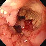
D- Know the endoscopic features of esophageal cancer:
In advanced cases, the appearance is generally very characteristic ulcers more or less tortuous to off-white background surrounded by a hard irregular bead in contact with the clamp, irregular bleeding and friable lesion vegetating blocked more or less light or impassable stricture.
Endoscopy is measured in all cases the distance between the upper pole of the neoplastic lesion and the mouth of Killian and search or permeation nodules or upstream dysplasia hearth of the tumor.
The limited lesions are more difficult to diagnose but should be well known because they allow the diagnosis to the stage of non-invasive cancer, the only form capable of healing.
It may be a mucosal range discreetly elevated or otherwise eroded, a simple change with frosted coloring and brilliance of the mucosa, or a small nodule.
It is always necessary to obtain a histological confirmation of several biopsies or smears made from elements of brushing an impassable stenosis.
E- Know the main staging elements and operability of esophageal cancer:
The loco-regional extension is sought by:
– ENT examination mainly looking for a recurrent reached and tracks concomitant neoplastic lesions of the upper airway.
– The tracheobronchial endoscopy (for cancers of the upper and middle third) for asserting an extension to the respiratory tree shows when an aspect vegetating or infiltrating or fistula, or just a simple tracheal or bronchial delivery which does not prejudge the parietal invasion.
– Assessing the parietal and mediastinal extension by CT vagueness when it comes to differentiating the simple touch of a tumor with a mediastinal organ and invasion of it.
The EUS allows more accurate assessment and better precision extension in different wall layers. It enables to distinguish purely submucosal lesions (Tl), lesions infiltrating the muscularis (T2), the mediastinal fat (T3) or adjacent organs (T4).
It can also indicate the existence of nodes but certainly affirm their cancerous invasion.
The lymphatic extension is sought by physical examination (ganglion Troisier).
Ultrasound Research cervical lymphadenopathy and celiacs and endoscopic ultrasonography to search for mediastinal lymphadenopathy are not practiced in all centers.
Visceral metastases are sought by a chest X-ray front and profile, liver ultrasound and possibly a chest and abdominal CT scan.
The synthesis of staging is currently done in the framework of the TNM classification.
The assessment of operability includes assessing:
– The nutritional impact, judged primarily on weight loss;
– Age;
– Liver function (TP);
– Respiratory function (EFR, unsystematic).
F- Know esophageal cancer of the principles of treatment:
The curative treatment is still currently based on surgical excision. Complete excision of the tumor must pass 5 cm above the upper pole of the tumor include the removal of the peri-tumor tissue and lymph node dissection.
Various surgical approaches are possible (with or without thoracotomy) and the replacement of the resected esophagus is usually achieved by means of a gastric tube.
Surgical treatment is only possible in patients able to support this intervention (age, general condition, respiratory, cardiac and hepatic function) and whose tumors do not include invasion of adjacent structures or metastases.
Preoperative chemoradiotherapy may be proposed because it increases resectability rates and even provides (in a third of cases) preoperative tumor sterilization and on parts of resection.
This result has not coincided with an improvement in survival.
The endoluminal radiotherapy and tumor destruction by laser photocoagulation may be considered as a curative treatment in the surface forms not exceeding submucosa (with a 10% risk to let evolve lymph node metastases).
Different therapeutic methods palliative aim to restore and maintain sufficient esophageal sector and prevent the establishment of a gastrostomy.
These methods are essentially endoscopic: endoscopic dilatation iterative, trans-tumor endoscopic intubation unblocking photocoagulation or electrocautery.
We can also undertake as palliative radiotherapy and / or chemotherapy in combination or not with an endoscopic method.
These suggestions are against if surgical indication or forms immediately and in highly developed recurrences.
They improve the quality of survival.
G- Know esophageal cancer prognosis based on its extension:
All forms, esophageal cancer survival is less than 10% at 5 years.
It depends on the parietal extension.
Lymph node involvement also plays an important role: 25% survival at 5 years in the absence and 10% 5-year survival in the presence of lymph node involvement.
In the presence of metastases (supraclavicular and celiac nodes are considered metastasis), the 5-year survival is zero.
You must be logged in to post a comment.