Generalized edema correspond to the accumulation of water in the interstitial fluids.
MECHANISM:
The passage of plasma water into the interstitial area depends on three factors:
– Transcapillary hydrostatic pressure which increases in case of venous or lymphatic obstruction;
– Osmotic pressure-related proteins (oncotic pressure) which tends to hold water in the plasma area and decreases with hypoalbuminemia;
capillary permeability to proteins which increases under the influence of vasoactive substances (histamine, bradykinin, etc.), derivatives of membrane phospholipids (prostaglandins, leukotrienes, platelet activating factor),cytokine (interleukin-2, granulocyte macrophage colony stimulating factor [ GM-CSF], vascular endothelial growth factor [VEGF], etc.) and complement components (C5-C9 fractions late or attack complex).
EXAMINATION, CLINICAL EXAMINATION:
Examination:
The examination should help:
– Specify the acute or chronic nature edema;
– Seek personal or family history of edema;
– Search for a trigger to circumstance (drug or food outlet, sting or insect bite, hormonal status in women, etc.).
Clinical examination:
The clinical examination must:
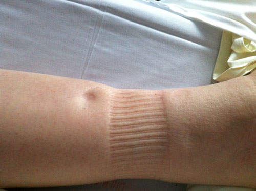
– Appreciate the importance of edema (one can earn up to 3 kg of water in the interstitial area without edema is visible);
– Seek urticarial lesions (puncture or otherwise in large erythematous and pruritic cupboards) or angioedema (swelling areas sudden skin or mucous membranes).
Full review:
The full review particularly interested four organs:
– The heart: find a r efl ux Hepatojugular;
– Liver: look for signs of portal hypertension and / or hepatocellular insufficiency;
– The kidneys: search proteinuria in the strip;
– Thyroid hypothyroidism but the hyperthyroidism may be generating edema.
Complete the review by the search for serous effusions. The reasoning depends essentially on the acute or chronic edema.
MEDICAL DIAGNOSIS AND TREATMENT:
Emergency treatment:
They are four in number, and fortunately rare.
Capillary leak syndrome iatrogenic:
Their Diagnosis is biologically the mismatch between blood count, which shows hemoconcentration (hematocrit> 50%), and protidogramme, showing hypoprotidemia by hypoalbuminemia.
A quasi-experimental capillary leak syndrome was observed in the treatment of some metastatic cancers (especially kidney) with high-dose interleukin-2 (IL-2) in combination with natural killer cells (NK) enabled the patient. This table is hardly observed with lower doses of IL-2 currently used, particularly those used in HIV patients to trace their CD4 count. There was a weight gain and peripheral edema, macular erythema and desquamation, peritoneal and pleural effusions. In the most severe forms occurred pulmonary edema, cardiac arrhythmias, hypotension, oliguria, impaired alertness.
Idiopathic capillary leak syndrome:
Rare, described in 1960 by Clarkson. It causes edema of the face and / or lower limbs that can be generalized, abdominal pain with or without diarrhea, and collapse with oliguria which may cause the patient’s death. Clinical difference with capillary leak due to interleukin-2 injections is the absence of pulmonary edema. This syndrome thus involves other cytokines such as vascular endothelial growth factor. Biological characteristic is the existence of a monoclonal gammopathy or benign, or as part of a myeloma. Sometimes there is a decrease in C3 or C4 complement fraction at the time of access.
Emergency treatment is based on the plasma volume (colloids, albumin, etc.) sometimes associated to pressor amines (dopamine).
Fluid replacement may be responsible for secondary complications (compartment syndrome, rhabdomyolysis).There are no known preventive treatment. We proposed a combination of aminophylline and terbutaline.
Edema hereditary angioneurotic:
The hereditary angioneurotic edema should be considered in a man or a young woman looking for a sudden surge of angioedema can achieve angioedema or even fatal laryngeal edema in the absence of emergency tracheotomy. It can also cause acute abdominal attacks repeatedly, afebrile (which eliminates periodic disease). The angioedema should then be discussed along with porphyria or poisoning.
This autosomal dominant disorder described in 1888 by Sir William Osler was reported by Virginia Donaldson in 1963 to an abnormality of the complement system. The fraction of complement C4 is lowered when accessed.
Must then be assayed inhibitor C1-esterase which is collapsed in the usual genetic forms.
However, the rate of C1 esterase inhibitor may be normal, or because it is a purely functional deficit, or because it is a woman whose angioedema outbreaks are triggered by the use of sex hormones.
Preventive treatment of access danazol (Danatrol®) to the loading dose of 600 mg daily. In women with normal C1 esterase inhibitor, progestins (norgestrienone) can be effective. If concern access, emergency treatment is injection concentrates C1 esterase inhibitor.
The angioedema may be acquired, mainly during the connective such as rheumatoid arthritis or systemic lupus erythematosus. Is implicated in this case anti-C1 esterase inhibitor autoantibodies.
Eclampsia:
Eclampsia occurs end of pregnancy. The warning signs are:
– High blood pressure (systolic ≥ 140 mmHg, diastolic ≥ 90 mmHg);
– Weight gain, p rotéinurie;
– An increase in serum uric acid is also a warning sign.
The primary risks are seizures.
A cesarean section should be decided in an emergency at the stage of pre-eclampsia. It should look for antiphospholipid antibodies in the mother postpartum.
Pushed acute edematous:
Acute edematous relapses are mainly drug and / or allergic.
Two specific etiologies must be evoked in women. Finally, the complete blood count and proteinuria used to find three rare etiologies.

Of generalized edema responsible medications:
The list grows each year (Table I).
Newcomers to the list are sartans (inhibitor of angiotensin II), glitazones (diabetes treatment and insulin resistance), imatinib mesilate (Glivec), which is a very effective kinase antityrosine in treatment of chronic myeloid leukemia or stromal tumors. We must add managers drugs drug related eosinophilia with systemic symptoms (DRESS), the most famous are the Disulone® (especially in the dark about), minocycline, allopurinol and certain anticonvulsants (carbamazepine mainly).
Drugs, we can relate a toxic, glycirrhizine licorice.Thereof, taken in large quantities can lead to greater weight gain of 3 kg and even authentic edema, through a hyperminéralocorticisme.
Indeed, glycirrhizine causes inhibition of 11-beta-dehydrogenase.
Food Allergies:
Food allergies can cause extensive acute edematous, localized to the face or diffuse, often associated with urticarial eruption. It is important to identify the food responsible, a later decision may cause anaphylactic shock. The fruits are large providers of food allergies, but curiously they hardly cause serious accidents.
In women of childbearing age:
The development of hydrops should be investigated taking inducers treatments ovulation (chorionic gonadotropin in particular). To avoid this sometimes severe ovarian hyperstimulation syndrome, it is imperative to supervise these treatments by repeat testing estradiol and ultrasound scanning.
Much more common, but less understood is the syndrome of idiopathic cyclic edema. After removing a plug occult diuretics or laxatives, patients often when going into orthostasis, hypovolemia, decreased urine output and sodium excretion. These defects are attributed to an increase of the capillary permeability of unknown origin.
Epinephrine orally would be effective in some patients.
Abnormal blood count:
If hemogram shows eosinophilia and increased immunoglobulin E (IgE) total, look firstly a trichinosis if a single thrust edematous with fever, second syndrome by Gleich. This comprises iterative surges angioedema, sometimes taking more than 5 kg of weight, eosinophilia greater than 1500 / .mu.l a hyperimmunoglobulinemia E, and also a steroid-hyperimmunoglobulinemia M. It is. It may be due to a clone of helper T cell TH2 (secreting interleukin-4 and interleukin-5).
Proteinuria:
The presence of proteinuria 0.5 g / 24 hours did evoke acute glomerulonephritis, especially if there is high blood pressure and an increase in serum creatinine.
Chronic generalized edema:
Chronic generalized edema should evoke many etiologies, among which it is possible to distinguish the causes should be suspected from the examination and clinical examination, and those that will highlight not only biology or after additional, sometimes complex investigations (Box 1)
Box 1. chronic generalized edema
Possible diagnosis from clinical examination
Pharmaceuticals
Chronic urinary retention (elderly)
Syndrome cellar
Endocrine:
Hypothyroidism
Hyperthyroidism
Diabetes (insulin-treated)
Heart failure
Constrictive pericarditis
Portal hypertension
Diagnosis require organic and / or complex investigations examinations
Hypoprotidemia
Cirrhosis
Nephrotic syndrome
Enteropathy
Infl ammatory syndrome
Kwashiorkor
Hypokalemia
Connective
Polymyositis and dermatomyositis
Granulomatous myositis
Diffuse systemic sclerosis
RS3PE
T-cell lymphomas
Yellow nail syndrome
Causes suspected from the clinical examination:
Medications:
The chronic edema drugs are mainly responsible calcium antagonists and glitazones.
Chronic urinary retention:
Urinary retention leads to edema mainly in the elderly with renal failure.
Vena cava syndrome:
The vena cava syndrome should be suspected in cases of thoracic collateral venous circulation and edema predominant in the supraclavicular regions (SVC syndrome can be difficult to distinguish from lipodystrophy Launois-Bensaude) or collateral venous circulation of the abdomen and buttocks (lower vena cava syndrome).
Endocrine:
Certain endocrine diseases must be mentioned at the outset, otherwise we risk ignoring the time. Edema predominant in pretibial regions are known in the myxedema. Less is known edema (up to hydrops) during major hyperthyroidism. The dosage of thyroid hormones confirms these assumptions.
Heart failure:
The diagnosis of heart failure in the presence of edema requires rigorous research Hepatojugular reflux, we should rather call abdominojugulaire test.
The patient should be in the semi-sitting position and breathe normally, and abdominal compression must be maintained for 10 seconds. Swelling of the jugular on operation may reflect right ventricular failure, pulmonary hypertension, or only an increase in pulmonary capillary pressure.
Constrictive pericarditis:
It is more difficult to make the diagnosis of constrictive pericarditis, which can be a cause of edema and ascites in particular. The third early diastolic sound is rarely individualized auscultation, echocardiography itself may be at fault, and you often go to the catheterization with fluid replacement to highlight the adiastolie. It remains to prove the origin of the pericardial adiastolie.
Portal hypertension:
Portal hypertension is suspected clinically if there is abdominal collateral circulation, especially if it is accompanied by signs of liver failure (many spider veins). The diagnosis is confirmed by gastroscopy shows esophageal varices and / or abdominal ultrasound study with the door system. In principle portal hypertension must be associated with hypoprotidemia to be a source of edema and ascites in particular.
Diagnosis requires further examination:
Hypoprotidemia:
The hypoproteinemia and hypoalbuminemia are mainly detected by electrophoresis of plasma proteins.
* Cirrhosis:
In France the leading cause of liver failure is Hyperalbuminemia, mainly due to alcoholic cirrhosis. (If there are no signs of liver failure and no signs of portal hypertension we must consider the Budd-Chiari syndrome and therefore request a study of hepatic veins pulsed Doppler ultrasound.)
* Nephrotic Syndrome:
Nephrotic syndrome is asserted if the serum protein is less than 60 g / L and there is proteinuria ≥ 3 g / 24 hours, knowing that kidney failure usually decreases proteinuria (except in cases where severe amyloidosis massive proteinuria persists despite the increase in serum creatinine).
* Protein losing enteropathy:
If the kidney and liver appear healthy, we are interested in the digestive tract and research hypertrophic gastritis Ménétrier by endoscopy or losing enteropathy. It can exist in the absence of diarrhea. Lymphopenia is a meaningful biological sign. The increase in fecal clearance of α-1-antitrypsin used to tell. The multiple causes of exudative enteropathy are recalled in Chapter chronic diarrhea.
* Inflammatory Disease:
Prolonged profound hypoalbuminemia, the inflammation may be sufficient to cause edema.
* Kwashiorkor:
In Africa, undernutrition in young children causes the spectacular Kwashiorkor.
Hypokalemia:
Biological routine exams should also include a chemistry panel for chronic hypokalaemia can be due to edema.
Connective:
* Dermatomyositis and polymyositis:
Among the connective dermatomyositis and polymyositis can cause generalized edema. The diagnosis is easy in case of dermatomyositis on the review of the integument (Liliace edema of the eyelids, fingers Gottron papules, erythema of the trunk). It is confirmed by capillaroscopy that shows megacapillaries.
The Creatine phosphokinase-(CPK) still increased in dermatomyositis and polymyositis with edema. The diagnosis is confirmed by muscle biopsy.
Granulomatous myositis:
Sometimes we are surprised to find on biopsy an aspect of granulomatous myositis whose main cause is sarcoidosis.
Systemic sclerosis:
Two forms of systemic sclerosis may
start with generalized edema.
One is relatively benign is Shulman’s syndrome, eosinophilia fasciitis.
This form of scleroderma is Raynaud’s phenomenon without and without visceral involvement.
Blood counts should be monitored because of eosinophilia and because this syndrome is sometimes complicated by myelosuppression making gravity. The other is serious is diffuse systemic sclerosis with Raynaud’s phenomenon, megacapillaries in capillaroscopy, visceral including renal crisis is the most serious.
RS3PE:
The RS3PE (relapsing seronegative synovitis with pitting edema symetrical) is an inflammatory disease of the elderly that can individualize or connect to a peripheral form of polymyalgia rheumatica.
T-cell lymphomas:
Revealed by edema, they are frequently accompanied by eosinophilia. The diagnosis can be focused on the study of circulating lymphocytes or biopsies (skin, muscle, lymph node) but sometimes biopsies are needed.
Yellow nail syndrome:
The yellow nail syndrome is exceptional, combining a lympho-edema, yellow nails and serous effusions on chest radiograph that allow diagnosis.
You must be logged in to post a comment.