The diagnosis is in many cases obvious. The patient consults because he complained mostly for the appearance of depigmented beaches on the skin.
The generalized pallor is observed in anemia, tissue perfusion decreases (vagal malaise, shock states …), the pan-hypopituitarism.
The hypomélanoses are characterized by a decrease in the amount of melanin present in the epidermis and hair follicles, whether due to a decrease in the number of melanocytes or a decrease in the synthesis of melanin. They represent the majority of cases of depigmentation. They are distinguished from non-melanoma depigmentations whose anemia is an example.
The causes of hypomélanoses are many but are easily recognized mostly by the examination (length of depigmentation, family history, comorbidities, local product applications and drug intake) by clinical examination and sometimes by a few simple laboratory tests (usually, thyroid function tests).
The hypomélanoses are mainly of origin:
– Genetic: albinismes oculocutanés, Piebaldism, mainly vitiligo;
– Endocrine: thyroid dysfunction, Addison’s disease, etc. ;
– Infectious: classically s secondary yphilis, tinea versicolor;
– Tumor: melanoma, nevus Sutton;
– After application of chemical agents;
– Post-inflammatory.
GENETIC HYPOMÉLANOSES:
Diffuse genetic Hypomélanoses:
They are easily recognized before a generalized depigmentation, up to the hair follicles, eyes, possibly with a family history.
In current practice, it is mostly albinismes oculocutanés.
Diffuse transmission is autosomal recessive genetic hypomélanoses. This is a heterogeneous group bonded to different genetic mutations. Risks are associated with photosensitivity and cutaneous neoplasia.
Diagnosis:
Clinical features skin depigmentation, high light sensitivity with the risk of skin cancer and eye damage. The skin is pinkish white, white hair, irises appear red in direct lighting in the most severe cases, often visible at birth. He is frequently strabismus, nystagmus, photophobia and decreased visual acuity severe.
In less severe cases, hair can be blond or light brown in life, with a few freckles and nevus achromic.
Irises are clear, blue, orange-yellow or light brown, with less loss of visual acuity. In the subject of black skin, the appearance is that of a blond caucasian.
For the record, some rare causes depigmentation give only a discrete, visible only when comparing the patient’s skin to that of family members. These pigment expansions, in particular linked to metabolic disorders (e.g., copper in Menkes disease, methionine in homocystinuria, phenylalanine in phenylketonuria).
Treatment:
Ophthalmological and dermatological monitoring is needed to treat and prevent complications. Photoprotection is applied.
Dyes hair or makeup may be necessary in case of aesthetic demand.
Early ophthalmologic care by a qualified optician with treatment of strabismus, concealers and tinted glass is required.

Genetic Hypomélanoses located:
Vitiligo:
These are mainly here of vitiligo, presenting clinically as an acquired lesion, but with a genetic substratum in over 30% of cases. It would affect 1% of the population.
It often appears in the second and third decades. Sun exposure often reveals lesions in contrast to the healthy areas taking tanning.
Diagnosis:
The clinical appearance is that of white blotches, although limited, confluent, localized, rarely generalized. Is the surface normal. The plates are sensitive, often starting on exposed areas.
The damage can be localized or generalized.
The most commonly affected areas are those discoveries, axillae, genitalia, bony prominences, areas and peri-orificial traumatized areas (equivalent Koebner syndrome).
The scalp (white lock), the bristles may be depigmented.
The evolution is unpredictable and capricious. Repigmentation may start with the hair.
It will look for arguments to an autoimmune disease associated diabetes, hyperthyroidism, or pernicious anemia or slow adrenal insufficiency.
The diagnosis is clinical. The etiological includes, in the absence of specific CIP, a blood count, a dosage of thyroid stimulating hormone (TSH) and thyroid antibodies Research (thyroglobulin antibodies and antithyroid antibodies), fasting glucose.
The psychosocial impact is often important.
Treatment:
The proposed treatments were multiple and often disappointing. UVB phototherapy (ultraviolet B) and narrow-spectrum laser are interesting.
Local corticosteroids can be used for localized lesions, but they face the risk of long-term complications.
Vitamin supplements and treatments for aesthetic purposes have also been proposed.
Waardenburg syndrome (or Piebaldism):
A white forelock achromic macules associated with the front of the chest and members evokes Waardenburg syndrome (or Piebaldism). There is also a group of heterogeneous genetic disorders. The syndrome may be associated with sensorineural hearing loss, widening the distance between the inner canthus and iris heterochromia. The mutation affects the region c-kit.
Tuberous sclerosis:
Hypopigmented spots in mountain ash leaves, lanceolate, associated with epilepsy suggestive of tuberous sclerosis.Angiofibromas of the face (nasolabial folds,
cheeks, forehead, chin, eyelids), fibroids peri-nail, skin plate lumbar pain, kidney tumors (angiomyolipoma, angiofibromas) and heart (rhabdomyomas) can join. Diagnosis is usually in childhood. Neurological involvement is often severe with mental retardation and behavioral disorders in addition to seizures.
The management is specialist.
Hamartoma achromique:
A hypopigmented macule, congenital, thorax and abdomen evokes a achromique hamartoma. No treatment is necessary.
Hypomelanosis Ito:
Depigmented lesions in splash in water jet, evoke a Hypomelanosis Ito.
This Hypomelanosis is genetic, related to mosaicism. Can be observed neurological disorders (mental retardation, seizure disorders, microcephaly), bone and eye damage.
Xeroderma pigmentosum:
Depigmented lesions, type of freckles or achromic macules few millimeters in diameter, can be observed in theXeroderma pigmentosum. It combine extreme photosensitivity, a motley appearance of the skin, poïkilodermique with multiple skin cancers (squamous and basal cell carcinomas), and melanoma, actinic keratoses.
Treatment is based on a medico-surgical management, with regular consultations to detect beginners carcinomas, resection of malignant lesions with cryotherapy or local destruction of 5-FU (Efudix®) actinic keratoses.Photoprotection (clothing, sunglasses, hats, long hair, photoprotective creams) while minimizing sun exposure is essential. Treatment with retinoids (0.5 to 2 mg / kg / day of isotretinoin) has also been proposed. Psychological support is needed. Prenatal testing is possible.
Other genetic disorders:
Some genetic disorders can be associated with pigmentation disorders: ataxia telangiectasia, Darier’s disease, von Recklinghausen’s disease, etc.
Canities can be observed in rare syndromes: progeria, Werner syndrome, myotonic dystrophy, Rothmund-Thomson syndrome, Fanconi syndrome, Fish, of Book, etc.
It can also be familial, with history found in the interrogation.
HYPOMÉLANOSES ENDOCRINE:
This is mainly vitiligo association and certain endocrinopathies:
– Diabetes;
– Thyroid dysfunction;
– Adrenal insuffi slow growth;
– Hypoparathyroidism;
– Chronic mucocutaneous candidiasis;
– Polyendocrinopathies.
Pigmentosa diffuse hypopigmentation is a sign of panhypopituitarism. Depigmentation of the skin of the genitals is observed in hypo-androgénisme.
HYPOMÉLANOSES TUMOR:
Vitiligo can be observed during the development of malignant melanoma.
Nevus Sutton is as nevus surrounded by a depigmented halo white.
Nevus may regress or disappear completely. This is normal during adolescence and young adulthood. In the older patient, an excisional biopsy is needed to avoid missing a malignant melanoma.
HYPOMÉLANOSES PHYSICAL AND CHEMICAL:
The examination found the notion of X-ray irradiation, exposure to ultraviolet (UV), burns, cold, mechanical attack.
Stopping physical aggression is necessary.
Depigmentation related to chemical agents can be professional, cosmetic, accidental or drug. The interview is crucial.
The majority of skin depigmentation is related to the application of topical treatments, in particular:
– Corticosteroids;
– Peroxides;
– Antimitotic in local applications.
Thio-uracil, used in the treatment of hyperthyroidism, has been incriminated. Mephenesin (Decontractyl®) butyrophenone, chloroquine give hair depigmentation.
Phenol derivatives are used in rubber industries, detergents, chemicals, disinfectants, printing inks.
Stopping the depigmenting agent may be necessary.
HYPOMÉLANOSES DEFICIENCY:
A Hypomelanosis can be observed during major denutrition:
– Kwashiorkor;
– Inflammatory bowel disease;
– Nephrotic syndrome;
– Small bowel resections important;
– Malabsorption, etc.
It particularly affects hair. The management of the underlying disease and nutrition is crucial.
HYPOMÉLANOSES INFECTIOUS:
Secondary syphilis:
Depigmented spots of the sides of the neck are a classic clinical signs of secondary syphilis (Venus necklace).
Onchocerciasis:
Onchocerciasis (river blindness) is observed in patients who stayed in tropical Africa, Central and South America and Yemen, especially in the course of rapid water’s edge. The hypopigmentation is related pruritus (itch filarial) particularly on the pretibial areas.
Kala-azar:
Kala azar is the visceral form of leishmaniasis (L. infantum, L. donovani). It can give pruritus related depigmented lesions is observed in patients of black Africa.
Pinta:
Pinta is treponematosis Central and South America. During the late phase are observed depigmented macules on the ends with pigmented follicular islets.
The processing is identical to syphilis.
Leprosy:
In leprosy, hypopigmented spots are conventionally squamous with disorders of opposite sensitivity. These are tuberculoid and indeterminate leprosy.
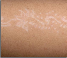
Tinea versicolor:
Pityriasis versicolor is a superficial fungal infection caused by Malassezia furfur. The hypopigmented macules are sharper after sun exposure. The lesions mainly affect the trunk and the root of the members. Initially, it is finely scaly maculae ranging from yellow to brown. Scratching is off dander (a sign of the chip).
Treatment is based on the application of an antifungal ketoconazole types (single-dose container Kétoderm® gel).We must apply the entire contents of a tube of gel (20 g) over the entire surface of the body including the scalp, lather and leave in place 5 to 10 minutes, then rinse thoroughly.
HYPOMÉLANOSES POSTINFLAMMATORY:
After an inflammatory skin disease, can be observed sequelae depigmented lesions.
The diagnosis is easily established by the interrogation. Can be observed and the depigmentation following the psoriasis, eczema, lupus, to insect bites, etc.
Depigmentation is more marked in black skin.
CAUSES OF VARIOUS HYPOMÉLANOSES:
Include the depigmentation observed in scleroderma, certain types of lichen, compared to nodules sarcoidosis, idiopathic gout Hypomelanosis (hypomélaniques small macules on the lower limbs of people sunning themselves), etc.
Syndrome Vogt-Koyanagi-Harada associated uveitis, aseptic meningitis, deafness, alopecia areata and vitiligo Hypomelanosis type. Hair and eyebrows are achieved. The patient is entrusted to the specialist.
Treatment is based on corticosteroids.
You must be logged in to post a comment.