The nails of the disease can be classified according to the clinical aspect and in the isolated or not the nail involvement:
– Coloring defects, shape, texture nails;
– Presence of a nail tumor;
– Nail disorders with the presence of another dermatosis.
The most common causes are infections, often favored by injuries (onychomycosis and paronychia candida).
The most formidable cause is acral melanoma, revealed by a nail brownish pigmentation (longitudinal melanonychia).
BACTERIAL AND FUNGAL INFECTIONS:
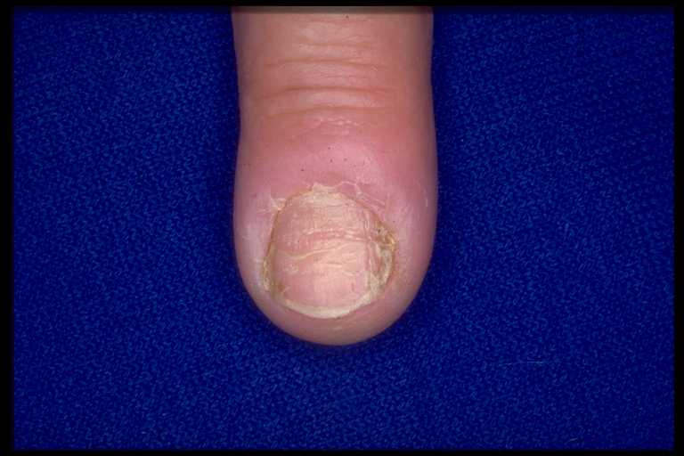
Reached nail:
It is a frequent pathology.
Diagnosis:
The nail is yellowish, thick and crumbly and sometimes even ends up destroying. A paronychia may be associated.The nails most commonly affected are those toes. Moisture, heat, strain and circulatory disorders are predisposing factors. A bacterial infection often plays a role: staphylococcus, streptococcus, gram-negative bacilli including Pseudomonas aeruginosa.
You have to practice a mycological sampling matrix if reached, several fingers or onycholysis before to initiate treatment. Sampling identifies usually a ringworm Trichophyton rubrum or type Trichophyton interdigital, a yeast typeCandida albicans. In difficult cases, it is the histology of the nail which rectifies the diagnosis.
Treatment:
Avoid contributing factors (tap ports in pools, treatment of footwear by applying Pevaryl® powder).
Local treatment:
Local treatment uses the varnish. Ciclopirox (varnishes Mycoster®) applies daily then after 3 to 4 weeks in two weekly applications. Amorolfine (Loceryl®) applies once a week. The treatment is long, 3 to 6 months in the hands of 6 to 12 months with big toes.
Chemical avulsion by bifonazole- Association urea (Amycor Onychoset®) can be proposed.
We must apply the ointment once a day on the nail reached, cover the whole nail and leave in place 24 hours under occlusive dressing. You must then remove the dressing, bathing the affected nail in warm water and remove the portion softened using the scraper provided. This operation is repeated every 24 hours until complete removal of pathological nail (1-3 weeks). It must then apply for two months bifonazole (Amycor® 1% cream).
General treatment:
General treatment is indicated in case of matrix involvement and / or more than two nails.
The simplest treatment is terbinafine (Lamisil®) at a dose of 250 mg / day for 6 weeks to 3 months fingers, 3 to 6 months to toe. Recall the cons-indication terbinafi only in patients with severe hepatic or renal impairment, risk of dysgeusia and the need to perform liver function tests before initiation of treatment.
Onycholysis pure candidiasis can be seen in the fingernails. It is necessary to cut the affected areas and apply an antifungal cream (eg, ketoconazole [cream Kétoderm®]) on the nail bed. Avoid contact with water and wear gloves for housework.
Superinfection with Pseudomonas aeruginosa is treated with twice-daily applications of sodium hypochlorite (Dakin®).

Paronychia:
Paronychia is an inflammatory disease of the periungual edge. The nail edge is red, inflamed and painful.
Infection with Staphylococcus aureus:
Acute paronychia with pus after pressure evokes a staph infection, wanted by bacteriological examination.
The treatment is medical first by antiseptic baths pluriquotidiens kind of hexamidine (Hexomédine®) and possibly systemic antibiotics for 5 days with pristinamycin (Pyostacine®) 3 g / day.
Candidiasis:
Chronic paronychia immediately evokes candidiasis. This usually occurs in domestic or confectioner. Inflammatory bead is slightly painful.
Candidiasis can also occur on a chronic paronychia or psoriasis.
Inflammatory chronic paronychia must also remove a malignant tumor and herpes.
We must protect hands by wearing gloves and making a treatment identical to the previous and a local antifungal (cf. onychomycosis).
YOU DIE:
If all dermatoses can not touch one finger, it is necessary in this case, however, suspected primarily a nail tumor.
Glomus tumor:
Bluish lesion of the nail bed painful, especially at the slightest touch, the suspicion of glomus tumor. There is erosion of the distal phalanx of the radiograph.
The treatment is surgical.
Exostosis nail:
Deformity of the nail in a young person, especially the big toe with painful outgrowth sign diagnosing exostosis nail confirmed by radiography. Walking can wake the pain.
The treatment is surgical.
Pyogenic granuloma:
The Pyogenic granuloma is an inflammatory granulation tissue succeeding trauma or an ingrown toenail. This is a red tumor, painless.
The treatment is surgical with pathological examination to eliminate an amelanotic melanoma or squamous cell carcinoma.
Pseudocyst mucoid:
A painless nodule causing longitudinal depression of the next tablet on the dorsal surface of the distal phalanx of the fingers evokes a mucoid pseudocysts.
The tumor is treated by surgery or intralesional injection of sclerosing. The mucoid pseudocysts may however reoffend.
Warty lesions:
Warty lesions, vegetating, hyperkeratotic evoke nail warts.
They are peri- or subungual.
We must cut out under local anesthesia tablet to release. Some propose to cut the wart with a scalpel and then rub the wart with dilute Bleomycin. Two to three weeks of daily fat dressings are then necessary to healing.
If warty lesions are chronic, Bowen’s disease is discussed and treatment is surgery, after pathological examination.
Fibroids peri-nail:
Peri-ungual fibromas of the tablet or lateral grooves evoke a tuberous sclerosis. They can also be isolated.
Malignancy:
A malignant tumor is suspected in:
– Destruction of the nail;
– An inflammatory chronic paronychia;
– Ulceration;
– A drawling pain;
– A keratotic closet;
– A monodactylique onycholysis.
A monodactylique onycholysis is also fear Bowen’s disease (squamous cell carcinoma in situ), squamous cell carcinoma or melanoma (see melanonychia).
A biopsy is required.
The treatment is surgical.
PROBLEMS OF COLOR NAIL:
Yellow nails:
Smoking is the most common and most obvious cause. Using external dyes, cosmetics and medicines (tetracyclines) may also be involved.
The yellow nail syndrome associated with yellow nails lymphedema, bronchopulmonary reached with recurrent pleural effusions, and possibly chronic sinusitis.
Brown fingernails or melanonychia:
A melanonychia is a nail pigmentation.
It may correspond to a lentigo, a nevus, a trauma, a fungal or bacterial infection, a foreign body, pregnancy, etc. The main fear is that of a melanoma.
Melanocytic hyperplasia:
Melanocytic hyperplasia is associated with either a benign proliferation (lentigo, nevus), or a malignant proliferation (melanoma).
A subungual black spot appears; it extends with the nail growth, gradually leading to the destruction of the nail bed.
Pigmentation of the nail edge (Hutchinson’s sign) is pathognomonic of melanoma.
A wide melanonychia with blurred edges, does not migrate with nail growth also raises melanoma. The nail melanoma can be amelanotic and look like a Pyogenic granuloma or a wart.
In subjects with darker skin, readily melanonychia affect several nails are mostly physiological (melanocytic hyperfunction). In the white matter, especially in case of single melanonychia, melanoma must be eliminated. In patients with dark skin, modification, or the appearance of a band melanonychia, especially in the fifth or sixth decade is suspect.
The patient is in all cases confi ed to the specialist.
Treatment after confi rmation pathological, is surgical, with wide amputation.
Melanocyte activation:
The melanocyte activation is responsible for the most commonly seen melanonychia.
The circumstances are different: pregnancy, hemochromatosis, Addison’s disease, trauma, Bowen’s disease, local carcinoma, taking cytotoxic, gold salts, antimalarials, silver nitrate, etc.
Repeated trauma:
Repeated trauma also cause melanonychia: onychotitillomanie thumbs with symmetrical involvement or involvement of toes rubbing in shoes.
Various causes:
Finally, there is occupational exposure to dyes, henna and abnormal pigment system.
White nails:
White nails (or leuconychia) are common.
The leuconychia is often due to abnormal keratinization of the nail. The most common cause is manicure, especially in women. More rarely, it is a Darier disease, autosomal dominant genetic disorder. Nail involvement is made of red and white stripes and distal subungual keratosis. There is no satisfactory treatment of this disease.
Congenital disorders can also give white nails.
White nails are also due to an abnormality of subungual tissue. The leuconychia then a call point to a general pathology (chronic liver failure in patients with cirrhosis, renal failure), dermatitis (psoriasis, lichen, eczema), taking drugs or toxic (sulfa drugs, chemotherapy, etc.)
TROUBLE THE FORM OF NAILS:
Clubbing:
Clubbing carries an expanded and curved nail watch glass (overcurvature nail).
The soft parts of the terminal phalanges can also be enlarged.
When acquired, bilateral, one must look primarily bronchial chronic suppurative (bronchiectasis), a lung tumor in a middle aged about smoking, a digestive pancreatic cancer especially cirrhosis and, rarely, hypothyroidism.
Unilateral forms should evoke an aortic aneurysm or the subclavian, abnormal brachial plexus, rarely aortic graft infection.
Clubbing is sometimes associated with hypertrophic osteoarthropathy pulmonary arthropathy Pierre-Marie.
There is no real treatment for clubbing. The possible cause should be investigated and treated.
Koilonychia:
Deformation nail spoon or koilonychia may be congenital or acquired. It is physiological in young children. Trauma, skin diseases, vitamin deficiency, hemochromatosis, polycythemia, infections are possible causes.
The treatment is the underlying cause.
Claw nails:
The claw toes are seen more frequently in big toe and in older women.
They are multifactorial: repetitive strain injury, fungal superinfection deficient circulatory status, neuropathy, wearing high heels, etc.
It must grind the nail reached then perform appropriate foot care. Wear proper footwear is required.
Large nails pincer nails:
The brachyonychie carries a wider nail than high. It is often congenital.
Nails clip can be congenital or acquired.
A nail clamp may be complicated by side incarnation of superinfection Pyogenic granuloma or the big toe.Hyperostosis of the phalangeal tuft is sought.
The treatment is surgical.
Compression in the shoes can cause pincer nails on the little toes.
The management is podiatry. Wear proper footwear is required.
The ingrown toenail usually occurs in young patients with nail plate a little wide and soft fleshy parts.
The treatment is surgical.
NAILS FRAGILE:
The nails are brittle, brittle, soft, with longitudinal hyperstriation.
It may be a physiological process related to the age and circumstances embrittling nail: trauma, immersion in hot water, detergents, solvents and sweet products. The eviction of these situations allows improvement.
It should also look for signs for a psoriasis, lichen or alopecia areata. Iron deficiency, sulfur, vitamins and medicines (retinoids) are also involved.
Treatment depends on the underlying cause.
In case of predisposing conditions, limit the maximum and wear gloves for manual activities, including the handling of vegetables. Nail care is to avoid, especially when performed in soapy water. The use of a moisturizing preparation and / or hardening of trade may be advised.
NAILS peeled:
The nail peeling is also called onycholysis. It corresponds to a mechanical cause a fungus, a psoriasis, an eczema, alopecia areata or a lichen. Handling corrosive or work in humidity are sought. A white onycholysis is often mechanical; yellow fungal or psoriatic onycholysis often. In this case, there is a subungual hyperkeratosis whitish, yellowish or orange color of the nail bed above onycholysis erythematosus and edging.
PROBLEMS OF ORIGIN TRAUMATIC:
Manicure:
Repetitive manicure with cuticles discharge, repeated soaking in water can cause paronychia, the leuconychia, transverse lines or onycholysis by passing the file under the shelf.
We must cut the protruding blades, avoid prolonged contact with water and stop manicure.
Onychotitillomanie:
Diagnosis:
The onychotitillomanie often affects inches.
Refoulement manic cuticle results in multiple transverse striations with a median longitudinal gutter appearance. The fingers used to repress the cuticle are often the index and middle fingers. Alternatives exist: the touch of a finger across the base of the thumb nail causing a median fissure, nail biting (especially children), paronychia by rubbing the pulp of a finger on the subungual fold, etc.
The large size of crescents is a marker of onychotitillomanie. A melanonychia friction can be observed. The gesture is sometimes unconscious on the part of the patient (tic), sometimes conscious (compulsion). Dermatitis underlying it sometimes associated with secondary onychotitillomanie.
Mechanical damage are also common to big toes. Microtrauma here favored by wearing high shoes, narrow, high heel, by acquired disorders of the foot and toes static (hallux valgus, overlapping toes, etc.)
Treatment:
Treatment of affected fingers is the prolonged wearing of occlusive dressings on the area. The patient must be aware of the tic.
For infringement of all fingers, one can propose the application of occlusive dressings rotatably (on some fingers first to cure, then others). Treatment with fluoxetine (Prozac) is sometimes necessary in severe cases.
If nail biting, applying topical (topical clindamycin, ketoconazole), unpalatable, is possible.
Subungual hematoma:
Subungual hematomas result from trauma, sometimes minimal and repetitive (as in the practice of hiking, toes, for example).
Support podiatry is essential.
Partial hematoma at the base of the nail, are treated by the perforation blade Lunularia region with a trombone red hot (lighter) or a scalpel. If major achievement of the tablet, the treatment is surgical.
Various violations:
Onycholysis 1 toe is sometimes caused by the overlap of the second toe on the first, longer than the first.Hyperkeratosis sousunguéale whitish can join.
The subungual horn is painful to pressure (walking, cloth) and is responsible for a distal onycholysis. It often affects the first toe. It is located under the shelf.
Her stripping relieves the patient. The bottom of podiatric treatment.
DERMATOSES REACHED WITH NAIL:
Psoriasis:
Nail involvement occurs in half of patients with psoriasis.
Diagnosis:
Surface irregularities are related to the tablet reached: nail pitting, roughness and tablet fragility. Leuconychia can observe. Onycholysis, subungual hyperkeratosis, a yellow stripe orange, salmon spots are also found. Nail psoriasis is often maintained by a Koebner phenomenon.
Depressions punctuated pitting are the most common. The roughness can lead to brittleness with tablet disintegration.
A greenish color may also be unconnected with Pseudomonas aeruginosa infection.
Treatment:
The patient is entrusted to the specialist. Treatment is difficult.
The nails are cut short, cut the peeled areas. Avoid repeated contact with water, soaking the hands in it.
The application of topical corticosteroids is degressive massage the nail bed in onycholyses. The attack treatment usually lasts 8 to 10 weeks.
Topical corticosteroids may be used under occlusion.
Delay shapes (Kenacort Retard®) can be injected into the subungual fold if matrix reached. Chemical avulsion urea hyperkeratosis may be required. If major and severe hyperkeratosis, treatment with oral retinoid (acitretin, Soriatane® 0.5 mg / kg / day) is proposed.
In the case of Acropustulosis, retinoids or methotrexate are shown in addition to the topical corticosteroid treatment.
Lichen:
The risk of the nail lichen is the healing process with destruction of the nail unit.
Diagnosis:
More than lichéniennes papules, achievement performs longitudinal striations and grésé appearance of the nails.Punctate erosions, onycholysis, subungual hyperkeratosis are also possible. The dorsal pterygium is suggestive: an extension of the cuticle extends cracking and destroying the blade.
Erosive lichen associates with nail lichen lesions post-bullous erosions.
Twenty nail dystrophy is seen in children.
The nails appear grésés. The evolution is spontaneously favorable. A lichénienne Nail involvement can also be seen in the disease graft against the host.
Treatment:
Treatment usually requires prolonged application of corticosteroids to prevent permanent scarring atrophy of nails.
Systemic corticosteroids may be required (prednisone, Cortancyl® 0.5 mg / kg / d tapering or monthly injection Kenacort Retard® 80 mg to 40 mg per month for 6 months). If unsuccessful, retinoids (Soriatane®) can be specified for a period of at least 6 months.
Eczema:
Eczema skin lesions are associated with nail involvement. The matrix is achieved, resulting in roughness, deformation, thickening and distortion of the blade. Onycholysis is possible. Beautiful lines or longitudinal striations may also occur.
Alopecia Areata:
Nail anomalies occur in a meaningful context with alopecia. Often observe grésés nails, punctuated erosions.
The fall of the nails is possible scopes and old forms.
No specific treatment is required.
The cure for alopecia areata causes improvement of nail lesions.
You must be logged in to post a comment.