The ulcers are losses of substance reaching the middle and deep corium with a risk of scarring.
The examination and clinical examination here have a major importance to reach the diagnosis.
There are basically acute injuries chronic injuries.

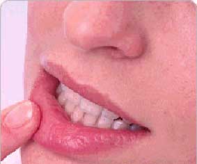
ULCERATIONS MOUTH:
The major forms are acute or recurrent ulcers and chronic ulcers dominated by the risk of cancer.
Acute or recurrent:
Aphte:
The clinical characteristics of ulceration associated with the sore are:
– Acute;
– Painful;
– Small;
– Yellow background surrounded by a red border.
Several clinical types are available: single, multiple, or only oral orogénitale (defining the bipolar aphthosis), necrotic or giant.
Benign idiopathic aphthosis:
The most common form is idiopathic benign aphthosis with a three elements of 3 to 4 mm in diameter, spontaneously evolving in 8 days to recovery, without scarring.
Outbreaks are rare with large asymptomatic periods.
Triggers are multiple, often different for each patient: food contact (nuts, cheese, chocolate), bites, stress, dental care.
No report is required.
Multiple aphthosis is defi ned by the presence of more than three canker sores.
Triggers are often the same, but the longer evolution (two to three weeks, with several waves).
Aphthosis miliary:
The aphthosis miliary is a special form of benign idiopathic aphthosis with 10 to 100 items, healing in ten days.Herpes infection must be eliminated (virological sampling).
Benign recurrent aphthous stomatitis:
It differs from the ordinary aphthosis by the number of relapses (more than four per year), sometimes subintrant. They can be punctuated by the rules (aphthosis Catamenial).
A general treatment is necessary.
Canker giants:
They are defined by the size of canker sores (one to three centimeters), often unique, necrotic or edematous. They meet especially among AIDS patients with a CD4 (cluster of differentiation 4) below.
Bipolar aphthosis:
It can be related to Behçet’s disease.
The following are then suggestive:
– Geographical origin: Mediterranean, Japan (actually in the path of the ancient Silk Road);
– Nature of the recurrent aphthous ulceration: more than three outbreaks each year;
– Skin involvement other than canker sores: erythema nodosum, pseudofolliculitis, acneiform lesions, hypersensitivity to puncture sites;
– Eye injuries: anterior uveitis with hypopyon, posterior uveitis, retinal vasculitis;
– Notion of recurrent superficial phlebitis;
– Possible neurological signs: hearing loss, vestibular dysfunction, cerebral venous thrombosis;
– Possible joint symptoms: arthralgia, mono- or oligoarthritis.
Diagnosis is also guided by the search for HLA B51 (human leukocyte antigen B51).
Other cases:
More rarely, aphthous ulcers, canker sores or real, can also be secondary to:
– Chronic intestinal bowel disease (Crohn’s, ulcerative colitis [UC]). The association with gastrointestinal symptoms is suggestive;
– Taking certain drugs (inhibitors of potassium channels such nicorandil);
– Neutropenia and granulocytoses occurring in a context of hematologic malignancy in the context of acute agranulocytosis iatrogenic or AIDS (possibility of giant ulcer).
The context and blood count (CBC) are evocative;
– The deficiency anemia (folate, iron, vitamin B12) often with ulcers on erythematous beaches;
– More rarely zinc deficiency (prolonged parenteral nutrition, malabsorption);
– Hypereosinophilic syndrome with skin lesions often hives, itching and high eosinophilia (> 1500), chronic (6 months), unexplained and visceral involvement (see pruritus);
– Cyclic neutropenia characterized by aphthosis beginning in childhood, cyclical (every 21 to 28 days) and sensitive to general antibiotic therapy.
Treatment:
Treatment consists of a share of common symptomatic measures in all types of aphthosis and also more specific treatments etiology.
Symptomatic measures:
They include:
– Antiseptic mouthwash with chlorhexidine (Eludril®);
– Mouthwash prednisolone (Solupred®) by melting 1-2 cp 20 mg in half a glass of water three to four times a day without swallowing, treatment which is associated a teaspoon of Eludril® ;
– Mouthwash by s ucralfate (Ulcar®) several times a day, diluted in a glass of water, then spitting the product;
– The application of Xylocaïne® viscous 2% before meals: 1 tablespoon to apply on lesions 3x / day (contreindiquées applications in porphyria, epilepsy uncontrolled by treatment and allergy to the product or the parabens; pay attention to the risk of choking);
– Mouthwash to aspirin (type Aspégic® 250 mg diluted in a glass of water in the quarterfinals then spitting out the product).
More specific treatments:
These treatments are:
– Eviction food triggers;
– Oral colchicine in the long term for recurrent oral aphthous ulceration and benign forms without visceral involvement of Behçet’s disease;
– Corticosteroids or immunosuppressants, for visceral involvement of Behçet’s disease that are the responsibility of specialist;
– Thalidomide in patients with severe aphthosis resistant to other treatments, to 100 to 200 mg dose for a month and then with progressive decrease. This teratogenic drug is subject to strict conditions of prescription and hospital delivery;
– Pentoxifylline (Torental®) at a dose of 300 mg / 3x / day that seems safe and effective.
Traumatic ulceration:
Clinical signs of traumatic ulceration:
– Single;
– Painful;
– In geographical contours;
– Necrotic
– After physical or chemical trauma.
In cases of inadequate dental prosthesis, dental trauma, contact with caustic, anamnesis easily identifies the traumatic ulceration.
The removal of the cause leading to their recovery in two weeks. Adverse changes made suspect the persistence of the cause, a tumor (required biopsy) or infectious complications.
Pathomimie:
Clinical signs of ulceration related to pathomimie:
– Single or multiple;
– Non-specific;
– In a particular psychological context;
– Do not fall within an identified semiotic framework;
– Without obvious cause.
The disorder is more common in the genital mucosa.
The management is delicate and must be based on psychiatry.
Herpes:
Oral herpes lesions are most often caused by herpes simplex virus type 1, but type 2 sufferers are rising.
Diagnosis:
In general practice, the diagnosis is clinical and requires no virological sampling.
Serology is of little use (only in case of primary infection herpes).
Always think of the potential severity of herpes infection in the person suffering from atopic dermatitis syndrome (KS-Juliusberg) and do not forget the possibility of immunodeficiency (HIV).
Herpetic primary infection:
Erosive febrile gingivostomatitis is for a primary herpes infection. This can be febrile, painful, hampering feeding, with satellite nodes. Primary infection rather occurs in children but may occur in adults.
Herpetic recurrence:
Clinical signs of erosion in favor of herpetic recurrence are:
– Painful or pruritic;
– Polycyclic;
– Succeeding a bouquet of vesicles on an erythematous base.
Herpes recurrences are easily identified by the patient himself. They recur at the same sites: lip with involvement of cutaneous side. Triggers are also well known patient fever, stress, sun exposure, menstruation.
Treatment:
In case of deep immunosuppression, treatment should be left to a specialist and usually in hospital (++ primary infection, acyclovir intravenously for ten days).
In case of symptomatic primary infection, if the oral route is not possible, and hospital treatment is based on the administration of intravenous aciclovir (Zovirax, 5 mg / kg / 3x / day for five to ten days). If the drug administration per os is possible, we give valaciclovir (Zelitrex®, 500 mg / 2x / day for ten days).
The treatment of oral herpes recurrence is difficult to codify. Topical antiviral drugs have not proved their effectiveness.
In minor forms, applying an antiseptic in aqueous solution like chlorhexidine and analgesics are sufficient. In case of frequent recurrences (more than six per year) and important, preventive treatment with oral valaciclovir (Zelitrex® 500 mg per os in a socket) can be offered.
In any case, research and adequate treatment of pain are necessary.
Preventive treatment for the entourage is based on simple hygiene measures (non-contact and non-herpetic lesions sharing machine).
Primary HIV infection:
Ulceration of the criteria associated with a primary HIV infection:
– Resemblance to the canker sore (aphtoïde);
– Morbilliform rash;
– Arthromyalgia;
– Headache;
– No obvious cause;
– Young with unsafe sex or drug addict.
Mouth ulcers may be associated with genital ulcers. The diagnosis is suspected on the data of the examination (risk factors) and confirmed by the p24 antigenemia.
The patient must be confi é a specialist in infectious diseases.
Other acute oral ulcers:
Zona:
Lesions similar to those of the herpes with
group on a dermatome should evoke shingles.
Infection with Coxsackie:
Vesicular pharyngitis with dysphagia or febrile combining oral vesicles oblong vesicles of the hands and feet should evoke an infection with Coxsackie (respectively herpangine piedsmains- and mouth syndrome). They are rather the prerogative of the child.
Treatment is purely symptomatic.
Secondary syphilis:
The depapillation in the language areas can be related to secondary syphilis (mowed plates).
Treatment is penicillin.
Anaerobic infection:
Ulcéronécrotiques of lesions, gangrenous in a subject debility (faulty oral hygiene, smoking, diabetes) or after non-steroidal anti-inflammatory (NSAIDs) should evoke an anaerobic infection.
The treatment is hospital with parenteral antibiotics covering anaerobes and based on surgery.
Sexually transmitted disease (STD):
Ulcers caused by STDs can be seen on the oral mucosa.
Erythema multiforme:
Mouth ulcers accompanied by bullous skin lesions and rosette must think of erythema multiforme (see, erythema).
Chronicles:
Autoimmune bullous dermatosis:
Clinical signs of erosion related to autoimmune bullous dermatosis:
– Chronic;
– Polycyclic or not contour;
– With possible damage to dental crowns;
– Combined with other bullous lesions on the seed coat.
Think especially pemphigus vulgaris and pemphigoid scar.
The standard histological biopsy and direct immunofluorescence, the search for autoantibodies confirm the diagnosis.
Squamous cell carcinoma:
Clinical signs of ulceration associated with squamous cell carcinoma are:
– Chronic;
– Painless;
– Indurated;
– Bleeding on contact;
– Favored by alcoolotabagique ground;
– With precancerous lesions keratosis;
– Erosive lichen;
– One or more lymph nodes and hard fixed satellites.
The patient should be referred to a specialist oral surgeon and otorhinolaryngological (ENT) (surgery, radiotherapy and chemotherapy).
Erosive Lichen:
Painful erosions with whitish network sign for a erosive lichen. Lesions can be cheek and lingual.
Chronic infection with hepatitis C and a risk of cancer should be conventionally sought.
The patient should be referred to a dermatologist. Treatment consists of topical steroids.
MUCOSAL ULCERATION GENITAL:
Think priority diseases (or infections) Sexually transmitted. Nevertheless, the same causes as those of the oral mucosa are present: mouth ulcers, infectious diseases, autoimmune bullous, trauma, cancers.
If the diagnosis of STDs is made, a systematic review is the rule with research other STDs including HIV prevention information, alert partner.
Genital treble:
Herpes:
Genital herpes can recover from a primary infection or be recurrent. In both cases, we must think about the severity of herpes in pregnant women (fetal and neonatal complications).
Herpetic primary infection:
Clinical signs of primary herpes infection are:
– Inflammatory erythema;
– Edema;
– Vesicles and erosions polycyclic in a context of feverish impaired general condition.
Primary infection can be extremely painful and cause urinary retention and vaginal discharge in women, and a urethral syndrome in humans. It typically occurs in young adults (for women).
It is most often due to herpes virus type 2. herpes skin lesions (vesicles, erosions) are possible in healthy skin.
Herpetic recurrence:
The clinical signs of a herpes recurrence are:
– Vesicles;
– Erosions grouped in clusters on erythematosus closet.
Genital herpes recurrences are often asymptomatic and raise the problem of the spread of the virus. The diagnosis is primarily clinical.
Virological sampling must be performed only in severe forms, especially in immunocompromised patients, during pregnancy and in atypical forms.
Treatment:
Treatment of primary infections and recurrences is identical to that of the oral forms.
In case of primary infection in pregnant women, if it occurs in the months preceding the birth, should be given aciclovir (Zovirax) at a dose of 200 mg / 5x / day orally until delivery ).
Pregnant women should be monitored by her gynecologist.
Condom use during and outside of relapses is recommended.
Syphilitic chancre:
Diagnosis:
Clinical signs of erosion due to a syphilitic chancre are:
– Own injury;
– Indurated;
– Although limited;
– Painless;
– Single (usually);
– Lymphadenopathy in the territory of drainage.
The syphilitic chancre occurs on average three weeks after infection at the point of penetration of Treponema. We must think about the possibility of secondary syphilis before papuloérosives genital lesions.
The diagnosis is confirmed by the positivity of the syphilis serology: FTA-abs (antibody [test] fluorescent treponemal)from the fifth day of the chancre, TPHA (Treponema pallidum complement fixation [test]) and VDRL (Venereal disease research labotary) to From the 7th day. HIV, viral hepatitis B and C and other STIs must be systematically sought.
Information on non-risky sexual behavior is required. Partners should be convened to break the contamination chain.
Treatment:
Treatment is based on the single intramuscular injection of penicillin delay (Extencilline®, 2.4 million units) in the absence of allergy.
In case of allergy, except in pregnant women, can be given doxycycline 100 mg / 2x / day orally for two weeks (risk of photosensitivity).
In pregnant women, treatment is the same unless there is an allergy to penicillin.
If necessary, the woman is entrusted to a specialist (desensitization before treatment).
Chancroid:
Diagnosis:
Clinical signs of ulceration associated with chancroid are:
– Multiple or not;
– Inflammatories;
– Non-indurated;
– Well-defined;
– Painful;
– Dirty background with purulent necrotic coating;
– Inflammatory satellite nodes.
Chancroid is caused by Haemophilus ducreyi.
He sits mostly on the skin side of the genitals. It appears approximately five days after the contagion. It is mainly related to prostitution (port or tropical contamination).
The germ is isolated on canker or lymphadenopathy.
Always seek associated syphilis.
Treatment:
The treatment is oral erythromycin
(Erythrocine®) at a dose of 2 g / day for 7 days or ceftriaxone (Rocéphine®) 250 mg in a single intramuscular dose.
Disease Nicolas Favre:
Clinically, signs of ulceration associated with Nicolas Favre disease are:
– Small size;
– Inguinal lymphadenopathy;
– Fistula.
Lymphogranuloma venereum is caused by Chlamydia trachomatis. It is rare in France.
Diagnosis is most commonly worn at the stage of multiple inguinal lymphadenopathy progressing to fistula. The germ is isolated from the pus of lymphadenopathy.
Serology is strongly positive.
Treatment is based on cyclins (oral doxycycline 200 mg / day or tetracycline 2 g / day for 21 days).
Fixed drug eruption:
A single lesion Bullous on erythematous background, a sign for a fixed drug eruption. It is rarely involved. The notion of prior drug intake is fundamental.
If réadmnistration the product, it tends to recur in the same place.
Taking the offending drug should be banned.
Erythema multiforme:
Erythema multiforme may also provide genital lesions associated with oral and skin lesions.
Treatment is covered in detail in Chapter bubbles .
Candidiasis:
Ulcers with inflammatory lesions covered with a whitish coating in the folds sign for a candidiasis. The land and history are suggestive (maceration, diabetes, antibiotics). The fungus is isolated from the mycological collection.
The treatment consists of topical antifungals application after washing (ketoconazole, kind Kétoderm®, one application per day for two weeks) and the management of contributory factors.
MST:
Ulcerations accompanying urethritis sign for a gonorrhea. We must think of others STDs, including syphilis (see Flows urethral).
Primary HIV infection:
Aphthoid ulcerations with rash and flu syndrome in young patients are in favor of primary HIV infection.
Chronicles:
Squamous cell carcinoma:
The first cause of chronic genital ulcer is squamous cell carcinoma. Any chronic genital ulcers should be biopsied for his research. As for the mouth, it can sit on a healthy mucosa, leucokératose, lichen sclerosus one, but also on Bowen’s disease.
Treatment is primarily surgical.
Other cases:
Extensive, chronic and painful ulcers in immunocompromised patients need to entrust the patient to a specialist. We must think of an opportunistic infection, tumor cause, atypical presentation of a viral infection (herpes, cytomégalvirus [CMV]).
You must be logged in to post a comment.