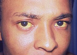
Jaundice is characterized by yellowing of the integument. It is most often visible at the top on the conjunctiva, the light area of the body wall. It is related to the increased plasma bilirubin and usually appears when this exceeds 40 mol / L. We will not deal here cholestasis of pregnancy or jaundice of the newborn.
DIAGNOSIS:
Faced with jaundice, the practitioner must eliminate an emergency (febrile jaundice, jaundice with liver failure, jaundice obstacle sudden onset) and determine the etiology, distinguishing between jaundice and bilirubin free bilirubin ones.
Before jaundice predominant free bilirubin, urine is clear and hemolysis is sought (NFS platelets, reticulocytes, LDH, haptoglobin).

Before a jaundice bilirubin, urine dark and cholestasis is sought (presence of pruritus clinically). In its presence, an imaging examination is performed to determine the presence or absence of an obstacle on the biliary tract. Jaundice with bilirubin are the most common, but cholestasis is only one of the causes of jaundice.
The presence of biliary colic type pain is an argument for a gallstone causing jaundice.
Other yellow colorations of the integument are exceptional and often foodborne.
The diagnostic approach is based first on the interrogation and clinical examination.
Examination:
The examination, in addition to standard questions, specifies in particular the surgical history of possible transfusion, hepatitis (especially in childhood, the patient often speaking of jaundice), tattooing, intravenous drug use, travel countries tropical.
An alteration of general condition and pruritus are also sought. An alteration of general condition and jaundice rather moving towards a neoplastic obstacle.
Biliary pain are also sought (epigastric pain, sometimes radiating to the right upper quadrant, precise beginning, gradually increasing to a phase plate, violent, in the absence of complications in less than twelve hours).
Fever succession evokes pain and jaundice cholangitis.
Family history of jaundice are also sought, as well as drug intake (not to mention the mode of contraception).
Ethnicity is also important: jaundice at free bilirubin by hemoglobinopathies among African or Mediterranean subject, greater frequency of hepatitis B and hepatocellular carcinoma in African and viral hepatitis in the Asian subject, etc.
Clinical examination:
It is comprehensive, focusing on abdominal examination.
Fever, itching, weight loss, hepatomegaly, splenomegaly, pain in the right hypochondrium and a ascites are sought.A big sign palpable gallbladder bile duct obstruction non gallstone (cancer, for example).
Additional tests:
The examinations to ask first line are organic and ultrasound.
Laboratory tests:
Are requested as first-line blood count, an activity assay of transaminases AST and ALT, gamma GT, alkaline phosphatase, total bilirubin and a dosage of TP, urea and creatinine .
The other reviews are based on the clinical context.
The arguments in favor of a hemolytic jaundice are the original character of unconjugated jaundice, decreased hemoglobin, reticulocytes rise, the fall of haptoglobin and the rise of LDH.
If cholestatic jaundice, it is bilirubin. There is then often pruritus, urine dark and alkaline phosphatase increased.
Imaging tests:
Doppler ultrasound:
In first intention, in jaundice in bilirubin, it is an abdominal Doppler ultrasound to apply for and obtain urgently.
This exam will determine if there is an obstacle in the extrahepatic bile duct, requiring surgical care or urgent endoscopic, visualizing dilated extrahepatic bile ducts. It also often helps to determine the nature of the obstacle (tumor, lymph nodes, etc.).
Cholelithiasis is observed on ultrasound by the presence of hyperechoic formations with posterior acoustic shadowing in the gallbladder or large bile ducts. Recall that gallstone disease is common in the general population and it is not necessarily involved in the pathology.
The headquarters of the dilated bile ducts to determine that of the obstacle, remembering that they expand over the obstacle. In case of obstruction of the convergence of hepatic ducts, intrahepatic bile ducts are dilated harmoniously.
In the event of obstruction on bile duct, the common bile duct is dilated. In case of bile duct obstruction, gallbladder and bile duct are dilated. If pancreatic obstacle, the bile duct and the common hepatic duct are dilated.
Ultrasound can more than look for signs in favor of cirrhosis (liver morphological abnormalities, ascites, signs of portal hypertension), cardiac liver (dilation of the hepatic veins and inferior vena cava) to examine the state of the venous system and hepatic portal, search for suspicious lesions, pancreatic especially cholelithiasis or deep lymph nodes.
Scanner:
The scanner provides identical elements on ultrasound but to better clarify the processes involved tissue (pancreas in particular chronic or pancreatic cancer, process invading the common bile duct).
It runs the risk of radiation and that of the contrast agent injection (renal failure, allergy).
Magnetic resonance imaging (MRI):
MRI dedicated to the biliary tract (bili-MRI) allows to visualize the bile duct and pancreatic duct. It is particularly interesting in case of suspicion of primary cancer of the biliary tract or primary sclerosing cholangitis.
EUS:
Endoscopic ultrasonography requires general anesthesia and therefore hospitalization. Preceded by a standard endoscopy, it allows the detailed exploration of the common bile duct and pancreatic region, particularly the lymph nodes.
It allows biopsies.
Cholangiography:
The ERCP requires general anesthesia. It runs the risk of acute pancreatitis and cholangitis. It can extract the calculations of the bile ducts, to carry out a sphincterotomy, ask a prosthesis.
URCENCES 1: JAUNDICE FEBRILE
Febrile jaundice within the emergency.
Jaundice free bilirubin:
Must move on the presence or absence of hemolysis (anemia, higher reticulocytes 120 giga / liter, elevated LDH, collapse haptoglobin).
Absence of hemolysis:
In the absence of hemolysis, detailed hereditary causes further (Gilbert syndrome and Crigler-Najjar) are mentioned.Intercurrent infection should be sought is the cause of decompensation of the underlying disease.
It should evoke mainly malaria (living in endemic areas, rarely vicinity of an international airport), abortion, pneumonia.
More rarely, the herpes group of viruses (CMV, EBV), hepatitis A and B, HIV are discussed. The babébiose is rare in France and provides an equivalent table to malaria.
Medications may be involved, by immunoallergic mechanism or G6PD deficiency (sulfonamides, amino-8-quinoline), beans.
We must not omit the possibility of autoimmune hemolytic anemia (Coombs test positive), ahead to seek the usual etiologies.
Chronic Hemolysis:
In chronic hemolysis, the dyserythropoiesis
(Disease Minkowski-Chauffard, hemoglobinopathies,
etc.) are sought. A
Again, just an intercurrent infection
destabilize the situation.
Jaundice bilirubin:
Cholestasis can be intra- or extrahepatic here.
The etiological investigation should be accompanied by a diagnosis of gravity, so as not to delay the treatment of urgent conditions (cholangitis, sepsis, fulminant hepatitis, etc.).
The research examined:
– The terms of the appearance of jaundice, speed of installation;
– The presence of an influenza-like syndrome, joint or muscle pain, headache;
– With impaired general condition;
– Abdominal pain, epigastric, hypochondriac, scapulars;
– A history of cholelithiasis or abdominal pain;
– Risk factors: foreign travel, freshwater swimming, occupation, recreation (fishing), drug addiction, alcoholism, transfusion history, taken medication, piercing, tattoos.
Dilated bile ducts:
Cholangitis:
The first diagnosis to evoke is that of cholangitis. Jaundice is by bilirubin.
The patient was admitted to emergency.
This is a biliary tract infection of bacterial origin. Sepsis is often associated primarily gram-negative bacilli, of intestinal origin.
Typical symptoms include succession in the order of right upper quadrant pain, fever and jaundice.
The first cholangitis occurs on a plot of stones in the common bile duct or large bile ducts. More rarely, cholangitis secondary to a prolonged non gallstone obstruction, such as cancer or primary sclerosing cholangitis.
Biologically, there is inflammation (neutrophilic leukocytosis, elevated CRP), cholestasis.
Blood cultures are performed, especially at the time of fever spikes. Renal failure is sought.
Ultrasound practiced emergency usually shows dilated bile ducts signing the obstacle. The pain in the right upper quadrant before the fever and jaundice evoke an obstacle, even if the bile ducts are not dilated at the time of the ultrasound.
* Treatment:
Emergency Hospitalization is required. The treatment is based primarily on the administration of intravenous antibiotics as soon as possible after completion of the bacteriological examinations, active in intestinal bacteria (usually Claforan® Association [3-6 g / d] + Flagyl® [ 3 × 500 mg / day] intravenously).
Hemodynamic disorders, kidney failure is possible to correct. The lifting of the obstacle is performed by endoscopic sphincterotomy in case of stones in the common bile duct, followed in a second time by cholecystectomy. In case of stenosis of bile ducts, a prosthesis can drain bile ducts.
* Differential diagnosis:
The differential diagnosis is that of cholestasis severe infections (pyelonephritis, pneumonia, peritonitis, typhoid, leptospirosis, etc.), severe acute alcoholic hepatitis, acute hepatitis A and E, of cholestasis sometimes observed during lymphomas (especially if there is a superimposed macrophage activation syndrome).
Other causes:
Neoplastic obstruction may cause jaundice by compression of the biliary tract, feverish or because of cholangitis superimposed, either because the tumor itself. The main tumors in question here are pancreatic adenocarcinoma, cholangiocarcinoma or vatérien ampulloma. Jaundice is progressive. There is often a big bladder and pruritus.
The patient is hospitalized in a specialized environment.
Parasites (flukes, roundworms), surgery or radiotherapy sequels are rarely involved.
Non dilated bile ducts:
Severe sepsis:
Severe sepsis may be accompanied by jaundice. Beware of septic shock, requiring transfer by ambulance in emergency resuscitation and emergency infusion of filling solution. The germs most often involved are gram-negative bacilli.
* Leptospirosis:
Febrile jaundice may be in the form of multiorgan icteric leptospirosis, most often due to Leptospira ictero- haemorraghiae.
Clinically, the 3rd day jaundice is observed classically influenza-like illness in a patient with risk factors (fresh water swimming, fishing, canoeing, sewage worker, veterinarian, fish farming, maintenance of water systems used , etc.).There is a jaundice bilirubin, often associated with kidney failure by tubulointerstitial nephritis.
Chest pain, hemoptysis, myocarditis or pericarditis may also occur. There may be a hemorrhagic syndrome associated with thrombocytopenia. It is feared septic shock, cardiogenic or respiratory distress syndrome in adults.
The patient must be hospitalized. The empirical antibiotic treatment is started in emergency. In the absence of renal failure, one can propose doxycycline at a dose of 200 mg / day. Other potential therapies are amoxicillin 100 mg / kg / day intravenously or ceftriaxone 1 g / day intravenously for 7 to 10 days. Complications are sought. The diagnosis is confirmed by serology, positive from the 8th / 10th day of evolution.
If exposed profession, he is an occupational disease. Preventive treatment is protection when in contact with potentially contaminated water (gloves, boots, goggles, overalls), protection of wounds and hygiene measures in case of wound (washing and antisepsis). There is a vaccination (vaccine Spirolept) to offer particularly exposed professions.
Serious infections salmonella, legionella, pneumococcus, mycobacterial diseases, brucellosis, Q fever, bartonelloses can also cause febrile jaundice conjugated bilirubin without bile duct dilatation and cytolysis.
Clostridium perfringens septicemia were seen after illegal abortions and are seen practically.
Thrombosis (portal, Budd-Chiari, rarely hepatic artery) can also cause fever and jaundice with conjugated bilirubin without bile duct dilatation.
Upon returning from a tropical country:
When returning from a tropical country, think first of malaria, especially Pl. falciparum and send the patient to the emergency. The babébiose can give a similar appearance. It should also evoke an arbovirus, including dengue and yellow fever (Africa and South America in unvaccinated subjects), for which hospitalization is necessary.
If the majority of cases are inconspicuous, the clinic can be noisy with high fever, headache, arthralgia and especially delayed hemorrhagic syndrome. Capillary leak syndrome may occur when dengue.
Yellow fever mainly affects America (Amazonia) and sub-Saharan Africa. Asia and the Middle East are spared. A vaccine is usually required for patients visiting these areas.
Clinically, severe acute hepatonephritis observed, hemorrhagic syndrome, encephalopathy (red congestive phase during the first days and then ictérohémorragique stage the following three days).
The diagnosis is confirmed by serology.
Treatment is symptomatic and preventive (fight against mosquito bites, vaccinations).
Hepatic amoebiasis (due to Entamoeba histolytica) may rarely cause fever and jaundice.
It occurs most often returned from an endemic country (Tropics, occasionally North Africa, exceptionally Aboriginal amebiasis).
There is usually pain in the right upper quadrant, rarely epigastric or left upper quadrant. Liver palpation is painful.Neutrophils and transaminases are high. There cholestasis.
Ultrasound reveals hypoechoic areas.
Serology is most often positive. The puncture is not necessary for diagnosis. It brings a chocolate-colored pus.
Treatment is metronidazole (Flagyl®) at a dose of 500 mg / 3x / day for ten days. Colonic home causing liver injury should be treated by a contact amebicide (tiliquinol-tilbroquinol, Intétrix®) at a dose of 2 capsules morning and night for ten days.
Acute alcoholic hepatitis:
Clinically, there are conventionally right upper quadrant pain, fever and jaundice. The minor forms are common, asymptomatic, resulting only by elevated aminotransferases. These minor forms lead to the development of alcoholic cirrhosis. The noisy classical form usually occurs in a subject already suffering from cirrhosis, alcohol poisoning after a major.
Depending on the severity of the underlying cirrhosis, ascites or encephalopathy can be observed. Bilirubin is high, to predominantly conjugated. The aminotransferases are high, the predominant elevation of aspartate aminotransferase. Alkaline phosphatase and gamma-glutamyl transpeptidase are usually high. There is a neutrophilic leukocytosis. Prothrombin time is generally lowered.
Ultrasound does not show dilated bile ducts. It can show a stéastose or abnormalities related to underlying cirrhosis.
Gravity is estimated by the score Maddrey (TP sick in seconds – TP witness in seconds) x 4.6 + Total Bilirubin (umol / L) / 17.
When this score is greater than or equal to 32, or if there encephalopathy, there is a severe acute alcoholic hepatitis requiring the production of a liver biopsy to confirm the diagnosis.
Hospitalization is required. Treatment is based on the power corticosteroids (Cortancyl®) 40 mg / day for 4 weeks in case of gravity.
Tumor process:
During observation of a tumor process in the liver ultrasound, various conditions can be observed.
* Malignant neoplasm:
A malignant tumor (primary or secondary), especially pancreatic adenocarcinoma, can be found.
* Hepatic pyogenic abscess:
One or pyogenic liver abscess, especially in diabetics or in case of infectious focus neighborhood, including abdominal, can be observed.
The distinction between abscess and liver tumor is not always easy.
We must hospitalize the patient to puncture, pus discharge with microbiological analysis.
* Hepatic amebiasis:
After returning from a tropical country.
* Hydatid cyst:
Liver hydatid cyst is due to Echinococcis granulosus. These cysts are observed especially in patients originating goats breeding areas (Mediterranean, parts of Africa, including North Africa).
Infection occurs after licking the hands parasitized by a dog, stroking the dog or contaminated food consumption.
It is most often latent, discovered incidentally.
Jaundice occurs when compression of the bile ducts. Cholangitis can occur in case of rupture of the cyst in the bile ducts.
Ultrasound or CT appearance is suggestive (cyst with parietal calcifications, presence of vesicles daughter, wall separation).
Serology is positive.
Do not puncture the cyst and typically assign the patient to a surgeon accustomed.
Surgical treatment is preceded by a treatment with albendazole (Zentel®) (15 mg / kg / day) daily for two months.
* Alveolar Echinocchose:
Alveolar échinocchose due to Echinococcus multilocularis, mainly affects patients who stayed in the East of France (Lorraine, Vosges, France County) and consumed wild berries contaminated by fox excrement or having skinned (hunters).
It carries a neighboring table of hepatocellular carcinoma, often revealed by hepatomegaly five to ten after the contagion. In addition to jaundice, pain in the right upper quadrant can be observed.
There are hypoechoic areas surrounding hyperechoic areas and dilated bile ducts on ultrasound. Liver macroscopic appearance evokes a malignant tumor.
Serology is positive.
Prognosis is poor. Partial hepatectomy is feasible when the parasite is located. Liver transplantation is sometimes considered.
* Malignant lymphomas:
Lymphomas Hodgkin and non-Hodgkin and certain lymphoid leukemia can cause jaundice by tumor infiltration or more rarely by compression of the common bile duct or cytokine production.
The liver failure is rare. Hepatic primary lymphoma is rare.
The patient is hospitalized in hematology entrusted to chemotherapy.
EMERGENCY 2: JAUNDICE WITH HEPATIC
Jaundice with liver failure within the emergency.
Clinically, are sought mainly for signs of hepatic encephalopathy (asterixis, confusion with reversal of circadian rhythm see coma). The spider nevi (more than five in the upper body, except in pregnant women), white nails, palmar erythrosis are in favor of a chronic liver failure. Haemorrhagic signs may be in connection with the decline in TP.
Biologically, liver failure is determined by the lower TP of factor V and albumin.
Acute liver failure:
This is most often acute hepatitis, viral (A, B, C, D, E, herpes), toxic (ammanite phalloides, paracetamol), drug immunoallergic, alcoholics. More rarely, it is an acute cardiac liver during a state of shock, for example. There may be an entanglement of different causes (eg, acute viral hepatitis worsened by the decision to phase préictérique acetaminophen or nonsteroidal anti-inflammatory drugs).
Encephalopathy is sought. The dosage of TP and Factor V is obtained urgently. The transaminase can be very high, greater than 20 times normal. The decline in PT and factor V less than 50% signed the severe hepatic impairment.The risk of severe hepatic impairment (encephalopathy) with a 80% mortality.
A low TP with normal Factor V signs the vitamin K deficiency, often related to cholestasis here. Intravenous administration of vitamin K then corrects hemostasis disorders.
Acute liver failure requires immediate hospitalization, usually by EMS, in specialized centers (hepatology resuscitation at best, necessary in case of reduction of factor V less than 50%, for a possible transplant).
Any paracetamol in this context, even at therapeutic doses, requires administration of acetylcysteine (Mucomyst®).An emergency liver transplant may be necessary.
In the liver cancer:
Jaundice occurs in advanced stages of primary or secondary liver cancer, often with liver failure.
The care is palliative.
Cirrhotic patients:
In a cirrhotic patient, worsening jaundice is seeking an aggravating factor:
– A push of the underlying causal disease: viral hepatitis, autoimmune, alcoholic, Wilson’s disease;
– An aggravating factor superimposed: hepatotoxic drug intake, gastrointestinal bleeding, especially infection of ascites, portal thrombosis, hepatocellular carcinoma, hemolysis on acanthocytes, worsening of renal failure, etc.
The patient is hospitalized, especially as it is also often an associated ascites thrust.
EMERGENCY 3: CHOLESTATIC JAUNDICE WITH VISIBLE EXPANSION OF BILIARY TRACT

Jaundice requires urgent specialist opinion with hospitalization (Fig 1).
It is due either to injury of the large biliary vessels viewable by imaging tests (ultrasound, CT, MRI-bili, EUS) or microscopic damage biliary vessels.
Recall that a prolonged cholestasis can cause weight loss by malabsorption but a progressive weight loss before jaundice is suspected of neoplastic cause.
If jaundice preceded by a biliary pain, most likely is that the obstacle prompt formation (eg gallstone migration).
Dilate the bile ducts above the obstacle.
For example, a large bladder signs choledochal obstruction and healthy bladder (otherwise the walls could distend).
Bile ducts may not expand upstream of an obstacle in case of multiple of their wall or hepatic fibrosis (cirrhosis). If recent constitution of obstacle, if the imaging examination is performed rapidly, the expansion can fail.
The obstruction of the common bile duct is a major cause of cholestatic jaundice with bile duct dilatation. The stones in the common bile duct is a cause. It can cause cholangitis and be even after cholecystectomy (residual stones).Pancreatic cancer and bile duct are also commonly involved. More rarely, compression of the bile duct by lymphadenopathy, with chronic pancreatitis calcification ante and postoperative stenosis of the bile duct can be observed.
Jaundice is late in case of primary biliary cirrhosis and must discuss liver transplantation. The mitochondrial type M2 antibodies are present, along with a rise in IgM.
More rarely, primary sclerosing cholangitis is a raised, often in a man with a history of Crohn’s disease or ulcerative colitis. Furthermore cholestatic icterus, it is the macroscopic appearance of the biliary tract which diagnosis: irregularity with alternating stenosis or expansions.
The occurrence of jaundice in a patient with sclerosing cholangitis is made evoke the development of cholangiocarcinoma, or a cholangitis.
CHOLESTATIC JAUNDICE NOT VISIBLE EXPANSION OF BILIARY TRACT:

It’s either a decrease or discontinuation of the biliary secretion, or obstruction or destruction of small intrahepatic bile ducts (Fig. 2).
Acute viral hepatitis:
Acute viral hepatitis caused by viruses A, B, C, D:
Recall that the acute viral hepatitis are asymptomatic in 80-90% of cases, and that the forms jaundiced only part of symptomatic cases.
Clinically, the symptoms are asthenia, headache, flu-like symptoms, abdominal pain with nausea. A fever can be seen as well as a rash.
Jaundice occurs secondarily, gradual onset, while the initial manifestations fade. The urine is dark, normal or pale stools.Jaundice gradually regresses in 2 to 6 weeks. Asthenia may persist.
In some cases, cholestasis is important (cholestatic forms).Encephalopathy may appear, accompanied by a decrease of TP or factor V less than 50%, defining fulminant hepatitis. The risk is 1% in case of icteric hepatitis B, 0.1% in case of icteric hepatitis A, lower in case of icteric hepatitis C. It is more than 1% in acute coinfection BD. The risk of death (90% if fulminant hepatitis B or BD, 50% if fulminant hepatitis A). The patient was transported by ambulance emergency resuscitation in hepatology in a center practicing liver transplant urgently.
The questioning search for specific risk factors for each virus: ingestion of water or food contaminated with feces, travel in endemic areas, homosexuals for hepatitis A; IV drug, surgery, transfusion, tattooing, piercing, blood exposure accidents (AES), unprotected sex for hepatitis B; IV drug use, AES, surgery to hepatitis C; Addiction, southern origin of Italy with Hepatitis D coinfection or superinfection BD; ingestion of drinking food or water contaminated by feces during a trip to Africa, South America or Asia for hepatitis E).
Biologically, transaminases are high, predominating in alanine aminotransferase. The gammaglutamyl transpeptidase is high between 5 and 10 N, normal or mildly elevated alkaline phosphatase. PT and factor V to be determined (severe or fulminant hepatitis if less than 50%).
Abdominal ultrasound is usually normal.
The serology to ask are: anti-HAV IgM the Hepatitis A; HBsAg, anti-HBc IgM antibody to hepatitis B, knowing that there is a 2 to 4 weeks window (the specialist will ask a search for viral DNA); anti-HDV antibodies to whether the patient has a known hepatitis B; anti-HCV antibodies with a window period 2 to 4 months.
According to the epidemiological context, an associated infection is sought (HIV, salmonella, typhoid, etc.)
Treatment:
There is no specific treatment, apart from severe or fulminant hepatitis and the patient should be transferred to emergency specialized Hepatology and hepatitis C where treatment can be provided by the specialist to limit the passage chronicity (pegylated interferon alpha 2a 180 mg / week with ribavarin 800 mg / day if weight less than 65 kg, 1000 mg between 65 and 80 kg, 1200 beyond). Alcohol and hepatotoxic drugs should be stopped, as all non-essential drugs. It will also remove the drugs metabolized by the liver.
A search of secondary cases entourage or source of contagion as the case must be made. Vaccination against hepatitis A or B must be done in the patient environment.
The main treatment is prevention (basic hygiene measures in endemic countries, vaccination for hepatitis A condom and vaccination for hepatitis B focusing infant vaccination, hygiene measures for hepatitis E) .
Herpes viral hepatitis Group:
Infectious mononucleosis:
Jaundice is rare in cases of infectious mononucleosis.
The diagnosis is supported by the ground (young adult), fever, sore throat, lymphadenopathy, the mononucleosis.Serology is to request the complete EBV serology test more than the MNI.
Cytomegalovirus infection:
The cytomegalovirus infection is rarely jaundiced (moderate fever for 3-6 weeks often hepatosplenomegaly). There can be mononucleosis; the IgM anti-CMV antibodies are present, without IgG. In uncomplicated cases of immunocompetent, there is no specific treatment to be started.
Chickenpox:
Jaundice is very rare during chickenpox.
Infections Herpes:
Infection with herpes simplex virus provide discrete jaundice in the disseminated forms, mainly in newborns, pregnant or immunocompromised.
The vesicular eruption is inconstant; vesicular rash is sought in the sexual partner or the mother to the newborn.
Fever and transaminases are often very high.
The patient is entrusted to the hospital, mortality is high, in order to start treatment with injectable aciclovir (Zovirax +) 15 mg / kg / 8 h IV.
Other viruses:
Include measles, rubella, echovirus, Coxsackie, paramyxoviruses, tropical viral infections, etc. Jaundice is fickle here, depending on the extent of hepatocyte necrosis.
Chronic viral hepatitis:
Transaminase elevation.
Autoimmune hepatitis:
They mainly affect women. They are chronic hepatitis but may be an acute jaundice in case of thrust.
There may be a family history of autoimmune hepatitis or personal autoimmune diseases: thyroid dysfunction, dry syndrome, vitiligo, etc.
The diagnosis is made by excluding other causes (especially HBV and HCV serology negative, the lack of antibody antimitochon dries) and the presence of autoantibodies in significant title: antinuclear factors and / or smooth antimuscle antibody specificity anti-actin on the one hand and on the other hand anti-LKM1 antibody (liver kidney microsome). More rarely, can be detected anticytosol of antibodies.
The patient is entrusted to specialist to immunosuppressive therapy (corticosteroids and azathioprine).
Toxic hepatitis:
Ammanite phalloides:
The poisoning especially ammanite phalloides occurs in the northern regions of France, Germany, Belgium and Switzerland, during the mushroom picking season (late summer, early fall). The examination finds the notion of consumer mushrooms picked in the forest, often family.
Jaundice occurs around the 3rd or 4th day after the onset of symptoms (abdominal pain, vomiting after a free interval of up to twelve hours and then elevated transaminases 2nd day). Liver failure may appear to the 6th / 7th day with encephalopathy (fulminant) with the possible death 48 hours later. Hepatitis can be less severe or even asymptomatic.
The patient was admitted to emergency intensive care unit in hepatology.
Paracetamol:
Jaundice occurs within 48 hours of taking a toxic dose of paracetamol (15 g in healthy subjects most often therapeutic doses in alcoholic malnourished, especially since he stopped drinking) . Liver failure with encephalopathy may appear between the 5th and the 10th day. The aminotransferases are often very high.
Treatment is based on administration of acetylcysteine as early as possible in hospital.
Other substances:
Other substances may cause acute toxic hepatitis, possibly jaundice: cocaine, ecstasy (hepatitis very cytolytic), paraquat, industrial solvents, plant (germandée small oak mistletoe, coal slime in Algeria, etc.)
Drug-induced hepatitis:
Clinical picture:
It may be in this case cholestatic hepatitis (alanine aminotransferase report on alkaline phosphatase expressed as the number of the normal less than 2) or cytolytic (ratio greater than 5).
If cholestatic hepatitis, cholestatic jaundice is with itching, dark urine, pale stools. There may be pain in the right upper quadrant and fever. Bilirubin is high, to predominantly conjugated with a significant cholestasis. The aminotransferases are moderately high.
The improvement is usually rapid and dramatic after stopping treatment. More rarely, the abnormalities may persist, in connection with a ductopenia.
If cytolytic hepatitis, in addition to jaundice, transaminases are very high, and gamma glutamyl transpeptidase alkaline phosphatase more moderately high or normal. The risk is the development of acute liver failure in connection with predominantly centrilobular necrosis.
The most common drugs involved are:
– Isoniazid (one to two months after the start of treatment, rather cytolytic hepatitis with risk of acute failure), especially since it is given combined with rifampin;
– Pyrazinamide;
– Ketoconazole;
– Sulfonamides (trimethoprim-sulfamethoxazole);
– Macrolides (cholestatic with fever, arthralgia, eosinophilia);
– Amoxicillin, alone or combined with clavulanic acid;
– Dextropropoxyphene;
– Non-steroidal anti-inflammatory;
– Halothane;
– Alpha-methyldopa;
– Captopril;
– Hydralazine, dihydralazine;
– The c arbamazépine, phenytoin, sodium valproate (microvesicular steatosis);
– Phenothiazines, t ricycliques, inhibitors of monoamine oxidase.
Diagnosis:
Diagnosis is based on the absence of serological markers for viral hepatitis, there is no obstacle on the bile ducts on ultrasound, a chronology of drug intake for the presence of immunoallergic signs (fever, arthralgia, eosinophilia), favorable after drug discontinuation (reintroduction is prohibited), the case described in the prior literature. If a liver biopsy was performed, it would show centrilobular necrosis.
Treatment:
Treatment is stopping or drugs involved and the failure to take any product nephrotoxic. In the most serious cases with fulminant liver failure, liver transplantation in super-emergency may be necessary.
Exceptionally, medications can give chronic hepatitis.
Severe Sepsis:
See sepsis.
Heart Liver:
This is usually more of a jaundice that a frank jaundice, predominantly conjugated bilirubin. Clinically, we can observe effort hepatalgia, firm hepatomegaly, a Hepatojugular reflux. Getting combine signs of heart failure: lower limb edema, jugular veins, oliguria, ascites.
The ultrasound shows dilation of hepatic veins, the inferior vena cava and hepatomegaly.
More rarely, cardiac liver takes the mask of acute or severe or fulminant hepatitis.
It is then usually acute heart failure (arrhythmias, acute pulmonary heart, pericardial tamponade see). The transaminases are strongly high (20 to 100 N), in case of healing of the causal disorder, rapid regression in 5 to 10 days.
Even more rarely, in cases of constrictive pericarditis or tricuspid lesions in particular, the evolution of cardiac liver can lead to the formation of an array equivalent to a true cirrhosis. The ultrasound shows dilation of the hepatic veins and inferior vena cava.
The treatment of cardiac liver is one of the underlying heart disease. The right ventricular failure is the most common cause.
The most common causes of chronic right ventricular failure are left ventricular failure, mitral stenosis and chronic pulmonary heart (hypoxic or postembolic), less often constrictive pericarditis. The most frequent causes of acute right ventricular failure are pulmonary embolism, right ventricular infarction, pericarditis, pneumothorax. Other causes are the tricuspid damage (secondary to right ventricular failure, carcinoid syndrome).
Various causes:
Primary biliary cirrhosis:
Jaundice is late in case of primary biliary cirrhosis and must discuss liver transplantation. The mitochondrial type M2 antibodies are present, along with a rise in IgM.
Cholangitis immunoallergic:
Medications can cause immunoallergic cholangitis with right upper quadrant pain, fever, eosinophilia. Macrolides, allopurinol, sulfonamides, amoxicillin-clavulanic acid are usually involved. Liver biopsy is needed.
Genetic Anomalies:
Even more rarely, have been implicated in the achievement of the bile ducts of small arms genetic defects (abnormalities MDR3 gene, cystic fibrosis).
Dubin-Johnson syndrome and Rotor:
Dubin-Johnson syndrome is rare, due to abnormal transport of organic anions.
Jaundice is fluctuating, often with a conjugated hyperbilirubinemia in predominance.
There are no cholestasis. The patient is entrusted to the hepatologist to diagnosis (liver biopsy). The disease is benign.
Rotor syndrome is another rare syndrome that is analogous.
IN JAUNDICE BILIRUBIN FREE:

The main causes are firstly hemolysis and dyserythropoiesis, and also the decrease in activity of bilirubin glucuronide transferase (Fig. 3).
Hematologic causes:
Hemolysis is diagnosed before a free bilirubin jaundice, macrocytic regenerative anemia, elevated LDH, collapse of haptoglobin, Coombs positive or schistocytes.
The dyserythropoiesis are treated in the chapter on anemia.
Recall hemolysis associated with valvular heart prostheses.
Hepatic Causes:
They are suspected in most cases in jaundice at free bilirubin (Gilbert syndrome) occurring in a young adult with a normal physical examination, after eliminating the usual big causes of jaundice, and with repeated history of jaundice and / or family jaundice.
Disease (or disorder) of Gilbert:
Disease (or disorder) of Gilbert is the most common of these conditions. Its transmission is autosomal recessive. An enzyme of the glycuroconjuguaison has a defective activity.
A superimposed hemolysis, fasting, intercurrent infectious episode may be the factor indicative of the disorder.Jaundice is moderate, often occurring in adolescence. Clinical examination is normal. Liver tests are normal, except for the free bilirubin elevation (often 20 to 80 mmol / L).
The disease is benign and most often requires no treatment.
Crigler-Najjar syndrome:
The syndrome Crigler-Najjar corresponds to a deficit of glycuroconjuguaison. The deficit is complete in type 1 which is a cause of neonatal jaundice in free bilirubin, a serious risk of encephalopathy. The defi cit is incomplete in the type 2, which has a better prognosis and requires taking a lifetime enzyme inducer phenobarbital type.
Benign recurrent cholestasis:
Benign recurrent cholestasis is rare, beginning before age 20, linked to an abnormality in the excretion of bile acids.It is autosomal recessive. Itching of jaundice occurs accompanied by episodes of weeks to months, yielding spontaneously.
The gammaglutamyl transpeptidase is usually normal while transaminases, alkaline phosphatase and bilirubin free are often high.
Treatment of episodes based on ursodiol (Ursolvan®, Delursan®). Rifampicin (Rifadine®) at a dose of 600 mg / day can relieve itching.
Intrahepatic cholestasis progressive family begins before the age of five years and are the domain of the specialist.
You must be logged in to post a comment.