Any breast lump should be explored in order to demonstrate its benignity. The physician should keep in mind the possibility of a malignant breast tumor then require taking prompt and specialized load.

CLINICAL REVIEW:
The context of discovery, age and history of the patient, the clinical aspect will allow diagnostic orientation but in the vast majority of cases, the prescription of radiological examinations will be necessary or even histological exploration.
Examination:
Context and background:
Family history (two ancestors achieved notably in pre menopausal, or association with ovarian cancer) may suggest the existence of genetic breast cancers: 5-10% of breast cancers linked to susceptibility genes (BRCA- 1, BRCA-2).
Personal history and mammary neoplasia risk factors should be noted (nulliparity, puberty before age 12 or late menopause, late first pregnancy …).
The date of onset and evolution over time should be recorded; appeared very quickly and a painful nodule is more in favor of a cyst or an inflammatory process.
Age:
The incidence of breast cancer increases greatly with age and while breast nodule appearing after 40, let alone after menopause is highly suspect and should be explored.
During the period of genital activity, the main diagnoses are:
– The fibroadenoma (tumor of the young woman 20-30 years);
– Fibrocystic breast disease (disease affecting women over 35 years);
– Breast abscess, especially during lactation.
Physical exam:
Clinical examination should specify (Table I):
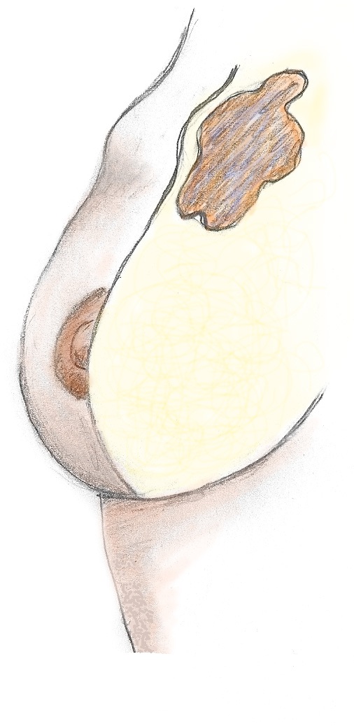
– The location of the nodule compared to the four quadrants of the breast;
– The size of the nodule;
– Consistency: elastic for a fibroadenoma, turned in favor of a fluid-filled cyst, hard and irregular for a malignant tumor;
– The limits of the nodule: Net in fibroadenoma and cyst, sometimes less clear in the case of cancer or very mastosique gland;
– Local inflammatory signs: breast abscess is a collection of pus rénitente, very painful, rather in young women, often during or after a feeding.
However, signs of inflammation may accompany cancer (ENP exacerbation 1, 2 and 3) thus forming an irregular mass pretty sharp contours with a possible appearance of orange peel. More rarely, it can be a granulomatous mastitis in the form of an inflammatory breast closet or still aseptic pseudo-abscesses;
– Mobility compared to skin, deep superficial layers: the fibroadenoma is mobile, the cysts are usually embedded within fibrocystic breast disease, cancers can invade the deep and superficial layers creating a skin deformation opposite either to type of retraction, or type of arch or deformation of breast contour;
– Mammelonnaire the appearance and the presence of a flow statements are beings: nipple retraction or discharge from the nipple sérosanglant unipore are suspect. Paget’s disease is eczematous skin lesions associated with mammary adenocarcinoma;
– Axillary lymph nodes are examined beings: the ipsilateral lymph node palpation nodule is suspicious, size and mobility of these should be recorded;
– In case of suspicion of advanced breast neoplasia, looking for secondary locations should be performed: bone pain, hepatomegaly, pleural effusions.

Additional tests:
Whatever the clinical appearance of the nodule, the discovery of it requires the prescription of additional examinations.
Mammography:
There are two pictures of each breast, an impact in the external oblique to identify good upper outer quadrant and inférointerne the deep planes and craniocaudal impact to visualize the surface part of the gland and the retro-areolar region. Additional copies can be made.
The radiologist will classify mammographic image RCA1 5 to guide the clinician on what to do (Table II).
Moreover, mammography allows the discovery of microcalcifications that are totally asymptomatic, frequent, variable meaning and difficult interpretation. The interest of these microcalcifications screening is to detect cancers at a stage when the disease is purely local. Classifying Le Gall is most commonly used to distinguish five types of microcalcifications according to their morphology, predictive value of increasing malignancy:
– I: ring or cup of tea (cup-shaped);
– II: Regular puncture;
– III: dusty;
– IV: irregular punctate;
– V: vermicular.
The number, distribution and shape of microcalcifications are important to enjoy.
If they are grouped, dense, irregular and number, they may be associated with cancer. Five suspected malignancy criteria are to remember: the worm-shaped microcalcifications, linear or tree layout, the size of the top home to 2 cm2, irregular shape and a number greater than 10 cm2 microcalcifications.
Usually microcalcifications do not sit in a palpable nodule; if their appearance evokes cancer, Mammotome biopsy or surgical excision after spear-tracking should be performed.
Breast ultrasound:
It complements mammography. During palpation of a nodule, it specifies the characteristics; it is more contributive when breasts are dense, especially among premenopausal women or in hormone replacement therapy.
If imaging is in favor of a cyst: rounded picture opaque in mammography, picture rounded anechoic fluid, a paracentesis may be proposed, relieving the patient and allowing a cytological study.
Histological examination:
The radiologist can during the exam, take samples for histological or cytological referred:
– FNA;
– Transcutaneous biopsy (true-cut) to confirm the malignant nature of the lesion and thus to organize surgical excision possibly associated with the technique of sentinel lymph node. For very large tumor, a biopsy will confirm and clarify the histology and neoadjuvant chemotherapy will be proposed to reduce the tumor volume for later surgery.
Histological control of certainty is sometimes necessary, several surgical techniques are available:
– Mammotome;
– Surgical excision, sometimes with prior radiological identification.

TREATMENT:
Suspected malignancy:
Any injury or ACR4 5 will be supported surgically for histological examination of certainty and tailor treatment.
The surgical management varies teams and the characteristics of the nodule.
Fibroadenoma:
If rapid change in the volume, echo
aesthetic or psychological, and after 40 years, surgical resection can be proposed.
In most cases, medical treatment allows at least partial involution of the lesion.
A antigonadotropic macroprogestatifs treatment can be offered:
– Lutenyl®: 1 tablet / day, 20 days out of 27;
– Orgametril®: 2 cp / d, from the 8th to 25th day of the cycle;
– Androcur®: 1 tablet / day, 20 days out of 27, in case of metabolic cons-indication or hyperandrogenism.
In the absence of surgical treatment, ultrasound and clinical monitoring is proposed at 3, 6 and 12 months to assess the evolution.
The hyperestrogenism climate favors the development of fibroadenomas and the combined oral contraceptives should be avoided.
Fibrocystic breast disease:
The ground hyperestrogenism should be avoided and can offer treatment by antigonadotropic macro progestogens (see above).
In case of painful cyst under pressure, paracentesis may be proposed.
Breast abscess:
An analgesic and anti-inflammatory treatment should be administered combined with systemic antibiotics. When the abscess is collected, surgical drainage should be performed.
You must be logged in to post a comment.