 Introduction:
Blood tissue distributed throughout the body, includes the hematopoietic organs and peripheral blood.
The peripheral blood behaves in two phases:
– A liquid phase, the plasma consisting of water, mineral salts, proteins, globulins with antibody activity, factors of hemostasis and nutritional factors.
– A cellular phase, consisting of figurative elements of the blood: red blood cells (red blood cells, erythrocytes), white blood cells (leukocytes), and blood platelets (thrombocytes).
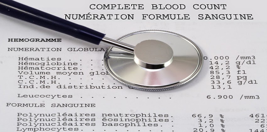
The hemogram includes: hematocrit measurement, hemoglobulin assay, red blood cell count, white cell count and platelet counts, calculation of globular indices (VGM, CCMH, TGMH) and calculation of other Parameters (automated leukocyte formula, Red Blood Cell Distribution Index (IDR), Platelet Distribution Index (PID), Platelet Volume (MPV).
The blood count must always be supplemented by the reticulocyte count and the study of the elements on a blood smear (FS).
The term NFS or FNS must be abandoned or even outlawed in medical language because of the limits of exploration of this examination (blood count and blood count).
Quantitative and qualitative study of red blood cells:
Red blood cells (RBCs), also known as red blood cells or erythrocytes, are anucleated cells whose daily production is mediated by erythropoiesis.
Their main role is to transport blood gases from the lungs (O2) to the tissues and tissues to the lungs (CO2) through hemoglobin.
The different methods of studying red blood cells are set out below:
Measurement of hematocrit (Hte):
The blood behaves like a suspension of elements figured in the plasma. The hematocrit measures the ratio of the volume (v) occupied by the figured elements to the total blood volume (V) of the analyzed blood sample. The unit of measurement is l / L or percentage (%).
The realization of a hematocrit rate requires the use of a dedicated microcentrifuge, adapted rods or capillary tubes, and modeling clay. The result is obtained after a centrifugation of 5 minutes at 1500 rpm.
The error on the measurement is plus or minus 2%; It is a reliable examination; It is simple and inexpensive. It is very useful in common medical practice to assess anemia. Normal values for age and sex are given in Table 1.
Red blood cell counts:
Two techniques are conventionally available to make a GR count: manual and automated.
1- Manual counting in Malassez cells:
The principle is based on the optical microscope count of GR contained in a given blood volume and known dilution; It is a method that is within the scope of any laboratory, therefore usable in all circumstances; This is unfortunately an unreliable method with errors of plus or minus 15% due to multiple causes (errors related to the operator, to the equipment used, or to the technique proper). It is a method that is now little used and replaced by automated measurement.
2- Automated counting by electronic meter:
It is a safer method but requires expensive equipment. However, it is currently available in most public or private analysis laboratories. The measurement error is + – 2% making this method very reliable. The measurements are usually carried out either by impedance measurement or by laser and more and more by flow cytometry. Normal values are given in Table 1.
Determination of hemoglobin (Hb):
It is made after transformation of the hemoglobin into cyanmethemoglobin: the optical density of the solution is measured at 540 nm by colorimetry; The measurement error is 10% in manual mode.
The pathological variations of the hemoglobin level are:
– Anemia: decreased Hb as a function of age and sex (Table 1).
– Polycythemia: Increased Hb level according to age and sex.
In true polyglobulia, Hb, hematocrit and rate of GR are increased in parallel.
Calculation of the globular indices:
The three measurements (Hte, GR, Hb) make it possible to calculate the VGM, the CCMH and the TGMH.
1- Average globular volume (VGM):
VGM = Hematocrit x 10 / number of GR in millions.
VGM = 80 – 100 Fl or μ3 (femtolitre or micron3).
This is the first important clue to characterize (type) anemia.
– If the VGM is less than 80 μ3, it is called microcytosis.
– If the VGM is between 80 and 100 μ3, we speak of normocytosis.
– If the VGM is greater than 100 μ3, it is called macrocytosis.
2- Mean corpuscular hemoglobin concentration (HCMC):
CCMH = Hemoglobin x 100 / Hematocrit. Unit: l / L or%.
CCMH = 32 – 38%.
This is the second important clue to characterize (type) anemia.
– If MHC between 32 and 38%, we speak of normochromy.
– If MHCC is less than 32%, it is called hypochromy.
– There is no hyperchromia.
3- Average hemoglobin globin content (TGMH):
TGMH = Hemoglobin × 10 / GR (million).
TGMH = 27 – 31 μg (picogram).
– If TGMH is between 27 and 31, we speak of normochromy.
– If TGMH less than 27, we speak of hypochromy.
The TGMH indicates the average hemoglobin weight in each GR; Its variation is parallel to that of the VGM: it is lowered in case of microcytosis and increased in case of macrocytosis.
Reticulocyte count:
Reticulocytes are young red blood cells that have just lost their nuclei in the bone marrow while continuing to mature for 24 to 48 hours; They then pass into the peripheral blood and become mature GR after 24 hours. They are recognizable after staining by brilliant cresyl blue or methylene blue. They reflect the quantitative daily production of red blood cells by the hematopoietic bone marrow.
The level of reticulocytes thus makes it possible to estimate the daily medullary production of red blood cells which is relative to 0.5 to 2%, ie 25,000 to 100,000 / μl / d. In practice, only the absolute rate is of interest. Thus, when the level of reticulocyte is less than 120 000 / μl, it translates a argenerative anemia (central cause). If the rate is greater than 120,000 / μl, it shows a regenerative anemia (peripheral cause) (Table N ° 1).
It is a key examination that specifies the central or peripheral mechanism of anemia.
Examination of red blood cells on blood smear (FS):
The blood smear is obtained after sampling and spreading a drop of blood on a glass slide; The smear is laid on a hard surface to dry spontaneously, after which it is colored at the May-Grünwald Giemsa (MGG). The reading of the FS is done with an optical microscope.
Examination of red blood cells under an optical microscope gives information on their size, color and shape; It is a very useful examination to characterize anemia: it confirms or invalidates the data obtained by the calculation of the globular indices.
– The main form anomalies are (Figure 1): anisocytosis, poykylocytosis, acanthocytosis, presence of target erythrocytes, dacryocytes, sickle cells, elliptocytes, schizocytes, spherocytes, and stomatocytes. The presence of RBCs = erythroblasts or punctate erythrocytes normally absent in peripheral blood.
– The size anomalies are: microcytosis (VGM less than 80 Fl), macrocytosis (VGM greater than 100 Fl).
– Color abnormalities are: hypochromia (HCMC less than 32%) corresponding to discoloration of the GR due to an insufficient concentration of intra-erythrocyte hemoglobin.
Quantitative and qualitative study of white blood cells:
The normal white blood cells of peripheral blood are represented by:
Neutrophilic or neutrophilic granulocytes (PN).
– Granulocytes Eosinophilic or polynuclear eosinophils (PE).
Basophilic granulocytes or polynuclear basophils (PB).
– Lymphocytes (L).
– Monocytes (M).
White blood cell count (GB):
The number of the GBs covers all the above-mentioned cells. The counting principle is the same as for the GR: manual and automated.
Manual counting is virtually abandoned in favor of more reliable electronic counting.
The rate of GB in adults is 4,000 – 10,000 / μl.
The leucocytic formula (leukocyte balance) or the respective count of the different GB categories. The result is given in percentage in the following order (PN, PE, PB, L, M).
The rigorous interpretation of the leukocyte balance is made on the absolute values calculated or given by the automaton.
The respective rates of the different GB categories are in adults (Table 1).
– PN = 40 – 70%, ie in absolute value: 1 600 – 7 000 / mm3.
– PE = 0 – 7%, or in absolute value: 0 – 700 / mm3.
– PB = 0 – 1.5%, or in absolute value: 0 – 150 / mm3.
L = 20-40%, in absolute value: 800-4000 / mm3.
– M = 3 – 7%, or in absolute value: 120 – 700 / mm3.
Pathological changes in white blood cells:
– GB greater than 10 000: hyperleucocytosis.
– PN greater than 7,000: neutrophilic polynucleosis.
– GB less than 4000: leukopenia.
– PN less than 1,600: neutropenia.
– PE greater than 700: hypereosinophil.
– PB greater than 150: hyperbasophilemia.
– L greater than 4,000: hyperlymphocytosis.
– L less than 800: lymphopenia.
– M greater than 700: hypermonocytosis.
– M less than 120: monocytopenia.
Examination of white blood cells on blood smear (FS):
Examination of the white blood cells at low and then at high magnification gives very useful information on their distribution (leukocyte balance), shape, appearance and size; It is a very useful examination to reveal certain abnormalities of the GB not given by the automata:
– Myelia: presence of immature granular cells (promyelocytes, myelocytes, metamyelocytes) normally absent from the blood.
– Presence of hyperbasophilic lymphocytes in viral infections (infectious mononucleosis) or parasites (toxoplasmosis).
– Presence of leucoblasts (blasts) revealing acute leukemia (myeloblasts or lymphoblasts).
The FS allows to establish an Arneth formula (FA) which is the evaluation of the distribution of the PNs according to the number of segments of their nucleus according to a curve (number of PN / number of segments); The right deviation (hyper segmentation of PN) of AF is seen in deficiency of anti-pernicious factors (vitamin B12 and folic acid).
Quantitative and qualitative study of blood platelets:
Blood platelets or thrombocytes are small sized elements from megakaryocytes of the bone marrow. They play a very important role in the mechanisms of hemostasis.
Platelet count:
Platelet count in Malassez cells or by automated counting is subject to a large margin of error (plus or minus 15%) requiring an indispensable visual control by blood smear before any thrombocytopenia. The normal platelet count varies from 150,000 – 400 / μl.
Examination of platelets on blood smear (FS):
The visualization of platelets on FS allows to evaluate their number and aspects (shape, size, granularity, clustered or isolated layout) in a fairly reliable way and thus to better interpret the data provided by electronic counting.
It is a very useful examination in the hands of an experienced cytologist.
On a SV made from a venous sampling under anticoagulant (EDTA: Ethylene diamine tetra acetic acid), the platelets appear isolated and distributed homogeneously on the slide.
On a sample taken from the finger, the platelets are found at the tail of smears in the form of clusters more or less important:
The presence of clusters of less than 5 platelets / clusters on the + side corresponds to a level of 50,000 to 100,000 platelets / μl.
The presence of clusters of 5 to 10 platelets / clusters on the ++ side corresponds to a level of 50,000 to 100,000 platelets / μl.
– The presence of clusters of more than 10 platelets / clusters on the +++ side corresponds to a platelet count greater than or equal to 100,000 platelets / μl.
One must appreciate the size of the platelets in search of macrothrombocytes often testifying to acquired or congenital thrombopathies, or rarely microthrombocytes.
At rates greater than 50,000 platelets / μl, patients do not bleed spontaneously, except in cases of local or general superadded causes.
Free thrombocytopenia (platelets below 50 000 / μl) and acute fractures easily lead to mucous, cutaneous or even visceral bleeding, it is hemorrhagic purpura.
The risk of severe haemorrhages, particularly cerebro-meningeal and retinal, is high below 20 000 platelets / μl.
Pathological changes in platelet count:
A level of platelets:
– Greater than 400 000 / μl characterizes thrombocytosis or hyperplaquettosis.
– Greater than 600 000 / μl defines thrombocythemia.
– Less than 150,000 / μl defines thrombocytopenia.
Overall variations of figurative elements of blood:
Bicytopenia: decrease of two bloodlines, eg anemia plus thrombocytopenia.
Pancytopenia: reduction of three bloodline lines, eg anemia plus neutropenia plus thrombocytopenia.
Cytopenias can be a peripheral mechanism (destruction, consumption, distribution) or central mechanism (defect of production).