Lymphadenopathy is an enlarged lymph node.
A node typically measures less than 1 cm; lymphadenopathy typically measure at least 2 cm.
Lymph nodes are lymphoid formations present on the lymph paths.
Their function is to destroy pathogenic (phagocytosis) and enable specific immune responses.
Lymph arrives in the node via an afferent lymph, flows through a lymphatic sinus network, then leaves the node via an efferent lymphatic.
The nodes are also vascularized (artery and vein).
The node structure includes the area around the hilum capsule, superficial cortex, deep cortex and medulla.
In the outer cortex, lymphocytes are organized into follicles. Primary follicles consist of unstimulated lymphocytes, whereas secondary follicles contain germinal centers appeared after antigenic stimulation. These germinal centers are the secondary site differentiation of B cells (antigen presenting cells: follicular dendritic cells).
The deep cortex is the induction site of the T-cell response (antigen presenting cells: interdigitating cells).
The medulla contains mainly macrophages and plasma cells.

DIAGNOSIS:
Clinic:
A superficial lymphadenopathy is diagnosed by palpation of lymph nodes. Superficial lymph nodes include the cervical area (pre and retro auricular, under chin, submandibular, occipital, cervical deep, superficial, and posterior), and in addition clavicular, axillary, épitrochléennes, inguinal. Sometimes it’s imagery that will highlight superficial or deep lymph nodes.
Before lymphadenopathy, interrogation will endeavor to find a notion of recent contagion, contact with animals (professional or family), travel, locoregional because of injury in the event of such a localized lymphadenopathy, impaired general condition, fever, night sweats, pruritus, arthralgia, tumor history, drug outlets, a notion of sicca syndrome …
The examination of lymph nodes must locate, measure, and describe the skin facing, consistency, mobility, sensitivity.
The full clinical examination must look for signs associated as hepatosplenomegaly, skin lesions, arthritis, neurological signs …
Additional tests:
Biology must include at least a complete blood count with blood smear (Research abnormal cells, hyperbasophilic lymphocytes), an inflammatory balance sheet (including a protein electrophoresis), liver function tests (typically disturbed in a viral context). Depending on the context, a lymphocyte phenotyping, an immunological assessment, a weight dosage of immunoglobulins, immunofixation, serology may be required.
Ultrasonography and CT can help to identify a necrotic or suppurative component.
The lymph node FNA can help to guide the diagnosis, unfortunately it is often non-contributory. It is interesting in the case of infectious adenitis because it may allow the identification of the germ.
The lymph node biopsy is the key consideration. The idea is to surgically remove the lymph node in its entirety. We must choose the largest ganglion and possibly avoid the inguinal area. The node is put in a fresh saline compresses; ganglion appositions slide are carried out; one part is fixed and will cuts for histological study, a party can be cultured, and another cryopreserved. Histological study allows in most cases a diagnosis.
In some cases, a thoraco-abdominal-pelvic imaging will be needed to view the other organs.
ETIOLOGY TREATMENT:
Infectious lymphadenopathy:
Localized lymphadenopathy (lymph node area):
Pyogenic adenitis:
* Mechanism:
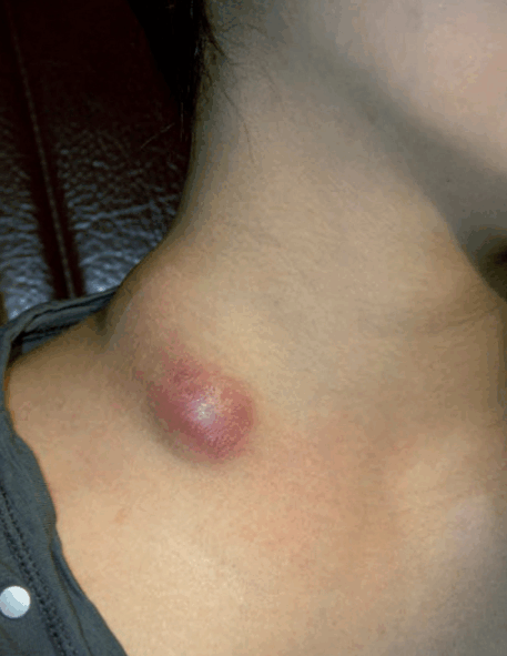
The lymphadenopathy is painful, causes fever and elevated neutrophils blood count; there may be a swelling in front of lymph node area or lymphangitis or perinodal cellulite. The germs are most often staphylococci or streptococci;the door can be a skin wound, oral or tonsillar infection.
* Treatment:
Treatment should include appropriate antibiotic therapy and sometimes requires an incision or paracentesis followed by drainage when lymph node is suppurating. Such adenitis, when they occur in children, are repeated, and are made of a gram-negative bacillus, have to suspect a chronic granulomatous disease associated with phagocytosis of polymorphonuclear deficit.
Mycobacteria:
The lymph node tuberculosis is the cause of lymphadenitis with coalescence on the same site; lymphadenopathy is initially firm and not painful, and soften, which adhere to the skin becomes inflamed, before fistuliser (“king’s evil”).The lymph node biopsy can show a tuberculoid granuloma with caseous necrosis, Ziehl positive staining (rare) and especially allows a cultivation that establishes the diagnosis. More recently, techniques of Polymerase Chain Reaction (PCR) have enabled faster diagnoses. General symptoms, the onset of erythema nodosum, the notion of contagion, ethnicity, immune status can refer to this etiology. Any tuberculous lymphadenopathy must seek another location and requires a tuberculosis treatment for 6 to 9 months.
Atypical mycobacteria can also give relatives tables; the diagnosis is the same conduct. Mycobacterium kansasii scrofulaceum and is mainly seen in children. Infections with Mycobacterium avium intracellulare are secondary to ingestion of unboiled raw milk and are seen mostly in HIV patients.
It should also be mentioned that combines BCGitis papular or ulcerated lesion of the vaccine site, and sometimes a satellite lymphadenopathy which may progress to suppuration and fistula.
The treatment is monotherapy with isoniazid.
Lymphoreticulosis benign inoculation or cat scratch disease:
The vector of this infection is Bartonnella henselae bacterium found in soil; inoculation is typically the result of a scratch or bite of a cat, but other animals such as rabbits and ferrets or simply spines may be responsible. Incubation lasts between 7 and 60 days; the presentation has a scar, an indurated papule or vésiculopustule at the site of inoculation, and painful satellite lymphadenopathy, sometimes bulky with little or no systemic signs.
This lymphadenopathy may persist for several months. She frequently progresses to suppuration and must then be punctured to prevent fistula; puncture brings a seemingly barren greenish yellow pus. The complications include meningoencephalitis, glomerulonephritis, pneumonitis and bone involvement. The diagnosis can be made through serology is positive if M or immunoglobulin G (IgG or IgM) are multiplied by four, or by culture and PCR of a lymph node biopsy. Lymph node histology is not specific showing granulomatous lesions and microabscesses; sometimes, the bacilli are seen in and outside the cells.
The treatment combines the puncture / excision of the ganglion and cyclin antibiotic therapy, fluoroquinolones, macrolides or rifampicin; the duration of treatment is not consensual, and ranges between fifteen and thirty days.
Pasteurellosis:
This infection is caused by a gram-negative bacillus, Pasteurella multocida. Infection occurs through the bite or scratch of animals such as cats, dogs and other mammals, but also plant sting. Incubation lasts forever under twenty four hours; the clinical picture is noisy, the inoculation wound becomes very painful, edematous, erythematous and well up leaving a serous fluid and pus. In one to two days appears satellite lymphadenopathy.
Early progresses to cellulitis, arthritis or sepsis if the patient is immunocompromised. Late complications are the result of subacute after remission of variable duration and are dominated by pictures of reflex sympathetic dystrophy.Diagnosis is made in the acute forms with the isolation of the organism in serous fluids and blood cultures, and in subacute forms by the concept of inoculation very painful wound.
The treatment of acute forms should last 10 days; the antibiotic of choice is doxycycline at a dose of 200 mg / day which may be substituted in children with amoxicillin or a macrolide. Forms sepsis are treated 15 days, with amoxicillin 100 mg / kg / day or doxycycline 300 mg / day. Unfortunately, in subacute forms, antibiotic therapy is no longer effective. Preventing pasteurelloses based on antibiotic prophylaxis wounds at risk by doxycycline 200 mg / day or 50 mg amoxicillin / kg / day for 5 days.
Plague:
The causative organism is a Gram-negative bacilli: Yersinia pestis. The vector is the chip that transmits the bacterium by stitching and reservoir consists of small rodents.
Plague is found in Asia, Russia, East Africa, Madagascar, South America and the western United States. Human transmission is via respiratory route.
Bubonic plague begins abruptly after incubation for 2 to 7 days; it is manifested by malaise, high fever and a toxic array (pallor, tachypnea). The review found one or more lymph nodes in a nodal site with local inflammatory signs.Direct examination of the aspirate is diagnostic highlighting the bacillus. The evolution in the absence of treatment is most often the death because of the spread of the germ or toxic syndrome. Pneumonic plague starts it, after incubation for a few hours to 3 days; the picture is that of a pneumonia with hemoptysis.
This form is very contagious and almost always leads to death despite antibiotic therapy. Diagnosis is made through blood cultures which highlight the germ.
Antibiotics called aminoglycosides, tetracyclines, quinolones, rifampin; the disease must be a national and international reporting. Antibiotic prophylaxis subjects refers to other tetracyclines or rifampicin.
Sexually transmitted infections (STIs):
* Primary syphilis:
Treponema pallidum is a spirochete that is transmitted in 95% of cases by sexual contact. Incubation lasts 3 weeks and appears superficial mucosal canker measuring 5-20 mm in diameter, although limited, painless, clean, accompanied by satellite lymphadenopathy (sometimes bilateral) non-inflammatory. Diagnosis can be made by demonstration of Treponema in dark field microscopy or serology positivation: FTA-Abs (Fluorescent treponemal Antibody Absorption) plus 3 weeks then TPHA (Treponema pallidum Hemagglutinations Assay) and VDRL(Venereal Disease Research Laboratory) positive.
The natural course is to the healing of canker in a few weeks with persistent induration while lymphadenopathy persists for several months. Treatment is to delay penicillins (Extencilline® 2.4 MU, intramuscular injection for primary syphilis of less than 1 year) or if allergic to macrolides and tetracyclines. The chancre disappears under treatment in 1-3 weeks.
* Chancroid:
This infection endemic in tropical regions is due to Haemiphilus ducreyi (Bacillus Ducrey). The short incubation (4-10 days); Clinical signs include a chancre or mucous, painful, dirty, non-indurated sometimes associated with inflammatory satellite lymphadenopathy which may progress to the fistula.
The diagnosis is established by identification of the bacillus in the sample by scraping the banks of the ulceration.The treatment of choice is a minute treatment with ceftriaxone 500 mg intramuscularly or Ciflox® 500 mg orally once daily.
* Disease Nicolas Favre or lymphogranuloma venereum:
This disease is also common in the tropics; it affects more male homosexuals (proctitis) in France. The germ involved is Chlamydia trachomatis. The infection progresses in three phases after incubation for 3 to 30 days: Initially, ulceration occurs not indurated, painless, then appear adenitis painful satellites, inflammatory which tend to coalesce may fistuliser; Finally, in the absence of treatment, onset of fibrosis with local secondary lymphatic insufficiency. The diagnosis is made by local cell culture sample, PCR on first urine stream or by serology which is always positive in this case. Treatment is to tetracyclines or macrolides for 21 days; if suppurative adenitis are, you have to avoid puncturing the fistulisation.
Note: reiterate the necessity for all STIs to detect and treat the patient partners.
Herpes virus:
Rarely, herpes or herpetic lesions can be accompanied by a satellite lymphadenopathy; these tables are seen more frequently in case of immunosuppression. Histology of the lymph nodes, which is not necessary, show foci of necrosis with cells at inclusion, witnessed the cytopathogenic effect of the virus.
Other infectious causes:
* Swine erysipelas:
The painting is dominated by a closet érysipélatoïde in the inoculation area in a patient handling meat or fish.
Complications are rare arthritis and bacteremia with endocarditis. Treatment is to penicillins.
* Tularemia:
This infection is transmitted to humans by handling rabbits (especially hunters).
The average incubation lasts 4 days and gives way to skin ulceration accompanied by satellite lymphadenopathy inflammatory. Without treatment, lymphadenopathy can fester for months. Diagnosis is made by serology. The treatment of choice is a double antibiotic therapy with cyclins and aminoglycosides.
* Sudoku:
The infection is caused Spirillum minus and transmitted by rat bite. Incubation lasts between 3 weeks and 2 months, and then show signs of inflammation at the bite scar with satellite lymphadenopathy, followed by the onset of fever, chills, headache, and finally a rash. In 2 to 3 days, the evolution is favorable but recurrences occur. The diagnosis is made by identification of the organism in dark field microscope from the removal of the skin wound, a node or blood cultures. Antibiotic therapy with penicillin or cyclins allows complete healing.
Differential diagnosis:
Two infections should be mentioned as they give pseudo lymphadenopathy:
* Donovanosis or inguinal granuloma:
Common disease in the tropics, it is due to a gram-negative bacillus, or bacillus Calymmatobacterium granulomatisDonovan. The incubation period varying from 1 week to 6 months which is followed by the appearance of one or more ulcers, indurated, granulomatous, painless, associated, not lymphadenopathy, but granulomas subcutaneous satellites that can coalesce and form a pseudobubon.
The diagnosis is made by culturing a sample by scraping the banks. Treatment with cyclins or cotrimoxazole to last 21 days.
* Actinomycosis and nocardiosis:
These gram-positive bacteria are the cause of soft tissue swelling pseudoganglionnaires and see themselves in patients with poor dental status. Diagnosis can be made by histology. Treatment is to penicillins if possible parenterally because of the fibrosis present in the lesions.
Polyadenopathies:
Viral:
* Infectious mononucleosis:
The table combines angina, cervical lymphadenopathy particular, moderate splenomegaly and mononucleosis in the blood picture. There are sporadic or extremely serious forms related Purtilo syndrome X-linked, which are often fatal and associated with hemophagocytic syndrome.
* HIV:
HIV infection is the cause of persistent polyadenopathies. We must therefore carry out a serology before a picture of polyadenopathy.
During primary infection with HIV, polyadenopathy joins a rash. However, it should not ignore HIV in a patient with lymphadenopathy other possible causes lymphadenopathy as tuberculosis, atypical mycobacterial infection, cytomegalovirus infection, lymphoma …
* Other:
Cytomegalovirus, adenovirus and rubella are also providers of lymphadenopathy. They are sometimes accompanied by a rash that is a guidance element.
Bacterial:
* Leprosy:
It is a cause of lymphadenopathy in endemic areas; they are then associated with skin lesions and peripheral neuropathy.
Hansen’s bacillus must be sought in the dermal juice lobules of the ear or skin lesions. The treatment combines at least rifampicin and dapsone or clofazimine for several years, with verification of the negativity of the sample.
* Tuberculosis:
See above (but generally relates to a lymph node area).
* Secondary Syphilis:
It may appear between the 2nd month and the 4th year of infection. It combines skin lesions (roseola, mucous patches, alopecia clearing, syphilitic) to polyadenopathy and sometimes hepatosplenomegaly. It is here that we find the classic épitrochléennes lymphadenopathy. The diagnosis is easily established by serology. Treatment should include three injections of 2.4 MU Extencilline® intramuscularly.
* Brucellosis:
The gram-negative bacillus infects goats and sheep; human contamination is most often professionals (veterinarians, shepherds, butchers …) but it can also relate to consumers of fresh dairy products.
Incubation lasts two weeks before the appearance of classical sudoroalgique fever that is accompanied by splenomegaly and sometimes hepatomegaly, and multiple lymph nodes.
The fever lasts a few days and then gradually decreases before rising again; these undulations are repeated.Biologically, there is a leukoneutropenia. Diagnosis is made through blood cultures and Wright serology. Treatment is associated with cyclin rifampicin for 6 weeks.
Whipple disease:
This is a chronic bacterial infection due to Tropheryma whipplei, which primarily affects males. Demonstrations conventionally include arthralgia, weight loss, central neurological disorders, digestive disorders but all organs may be affected. Lymph nodes are reported in 50% of cases. Histology whether lymph node, or other duodenal shows a foamy macrophages infiltrate by containing PAS positive material. The research Tropheryma whipplei by PCR can confirm the diagnosis; it must also be made on blood and cerebrospinal fluid. The treatment uses a parenteral initial combination therapy for 14 days with ceftriaxone 2 g / day and streptomycin 1 g / d and a relay Bactrim forte® 2 cp / day for 1-2 years. At the end of treatment, an assessment must be made to ensure the disappearance of the bacteria.Despite this treatment, relapses are possible. Patients should be monitored for life.
Parasitic:
* Toxoplasmosis:
The picture is that of persistent lymphadenopathy sometimes with mononucleosis in the blood picture. Serology allows diagnosis by showing the appearance of IgM and IgG. The only treatment whose effectiveness has been proven and is Malocide® Adiazine® association, which is prescribed only in severe forms of immunocompetent and in the immunocompromised. Treatment with 3 MU Rovamycine® / 3x / day can be discussed in symptomatic forms of immunocompetent, although its effectiveness has not been proven. Toxoplasmosis is also dangerous to pregnant women because of the risk of congenital toxoplasmosis; the risk of passing the fetus is more important from quarter to quarter; the consequences can be fetal deaths, congenital encephalitis, chorioretinitis, mental retardation … A pregnant woman who is a primary infection with Toxoplasma gondii will be Rovamycine® in order to reduce the risk of transmission of the parasite to the fetus; Search congenital toxoplasmosis and therapeutic management are made in obstetrical environment.
* Visceral Leishmaniasis:
This infection is present in Asia, Africa and South America. The parasite is transmitted by the bite of a sandfly, follows an incubation period of one to six months. At the status phase, the picture is dominated by disarticulated fever, pallor, weight loss, hepatosplenomegaly and painless lymphadenopathy. Biology shows anemia, leukopenia, thrombocytopenia often a significant hypergammaglobulinemia. The diagnosis is confirmed by the detection of the parasite in lymph node or bone marrow aspiration and serology.
* Trypanosomiasis:
Sleeping sickness is caused by a protozoan that is transmitted to humans by the bite of a tsetse fly. This infection is rampant in black Africa. After a variable incubation appears disjointed fever, lymphadenopathy, hepatosplenomegaly and sometimes polycyclic closets skin, headache and behavioral changes. The intracerebral reached then continues to evolve, leading to deficiency states and coma.
Biologically, the presence of anemia, a circulating plasma cells, hypergammaglobulinemia and a major elevation of IgM. The diagnosis is confirmed by the detection of the parasite in the blood, bone marrow, lymph node puncture juice, cerebrospinal fluid. In some cases, the serodiagnosis may help. The treatment is hospital, depending on the parasite and the disease phase (pentamidine, melarsoprol, suramin).
Fungal (histoplasmosis):
Histoplasmosis is a fungal infection import.
Two germs are distinguished: Histoplasma capsulatum is found in the United States, South America, Guyana, the Caribbean, Africa, India, and Histoplasma duboisii present in black Africa. Histoplasmosis Histoplasma capsulatumto initially manifested by a primary infection may be silent or give influenza-like illness with cough and dyspnea;imaging shows a miliary pulmonary and mediastinal lymphadenopathy.
These symptoms disappear within a few weeks leaving calcified pulmonary, hepatic and splenic scars. In immunocompromised patients, HIV, children and sporadic cases, histoplasmosis can spread with multiple organ damage (neurological, endocardial, adrenal, digestive, glandular). Histoplasmosis H. duboisii manifested by cutaneous lesions, bone or lymph node (in this form, localized lymphadenopathy). Diagnosis is based on the notion of living, even ancient, in endemic areas, the identification of yeast cultures (sputum, blood culture, pus, smears ulcerations, skin biopsy …), giant cell granuloma lesions in Histology of a lesion and a positive serology (but insufficient sensitivity). Treatment is to hold anti-fungal.
Proliferative pathology:
Hematologic:
Malignant lymphoid diseases:
Non-Hodgkin’s Lymphomas, Hodgkin’s disease, certain chronic lymphocytic leukemia and acute lymphoblastic leukemia manifested by lymphadenopathy. This is most often polyadenopathy that can be symmetrical or asymmetrical, be accompanied by hepatosplenomegaly, fever, impaired general condition with rapid and substantial weight loss, night sweats, pruritus .
Some clinical signs can point to the type of lymphoma. The painful nature of lymph nodes in the absorption of alcohol should suggest Hodgkin’s disease; the combination of joint and skin symptoms should suggest a T lymphoma; the combination of a rash, fever, lymphadenopathy and a poor general condition in an elderly patient should suggest angioimmunoblastic lymphadenopathy.
Biologically, there may cytopenias associated with bone marrow infiltration, eosinophilia, atypical lymphocytes in blood smear, a monoclonal peak in the case of a disease Waldenstrom.
The diagnosis is established through lymph node biopsy which will also typer lymphoma and so decide the treatment.
Angiofollicular lymphoid hyperplasia Castleman:
There are two forms of this disease: a localized form and a multicenter form. The localized form typically affects young adults and is characterized by a large localized lymphadenopathy, often mediastinal or abdominal.
The histology of resection material allows diagnosis.
The treatment of this form is surgical.
The multicentric form is more rare, concerns elderly or immunocompromised individuals and is characterized by general signs, polyadenopathy superfi cial. The change is more severe with a risk of multiple organ failure mediated by inflammatory cytokines, and Non-Hodgkin lymphoma.
Histology also allows the diagnosis. The treatment of these forms involves cytotoxic, anti-CD20, to thalidomide.HHV8 is involved in the pathogenesis.
Myeloid malignancies:
In some cases, there may be lymphadenopathy: in the case of acute myeloid leukemia, lymph nodes can be present in particular in the form myelomonocytic and monoblastic; in the case of myeloproliferative disorders, there is rarely lymphadenopathy associated with myeloid metaplasia nodes. The blood smear, complete blood count, myelogram, bone marrow biopsy allows the diagnosis in these cases.
Granulocytic sarcoma:
This is the primitive node location of acute leukemia. Diagnosis is made through lymph node biopsy and the blade on apposition.
POEMS:
This acronym signifi e Polyneuropathy, organomegaly, endocrinopathy, Monoclonal component, Skin changes.Lymphadenopathy can be observed in this pathology sometimes histology of lymphoid hyperplasia angiofollicular plasma cell types (multicentric Castleman).
Mastocytosis:
They are defi ned by the presence of too many mast cells tissue. They can be pure cutaneous or systemic. The symptoms are related to mast cell degranulation (flushing, hives, diarrhea …) and tissue infiltration (urticaria pigmentosa, splenomegaly, lymphadenopathy …).
The lymph nodes are encountered in systemic forms and are generally deep lymph nodes. Diagnosis is made on the histology of affected tissue (skin, gut …). The initial assessment should include a bone marrow biopsy and bone balance (BMD, bone scan) to determine the prognosis, some forms may be associated with blood diseases even more rarely catastrophic prognosis leukemia mast cells.
Treatment aims to prevent episodes of degranulation (regime against-indication of some drugs) and limiting symptoms (antihistamines, corticosteroids short) in indolent forms. In forms associated with blood diseases, chemotherapy are proposed; bone involvement is improved by the use of bisphosphonates; Finally, the effectiveness of interferon α has been shown in some cases of mastocytosis with major bone marrow infiltration. The revolution in the treatment of mastocytosis is the use of imatinib mesylate inhibits the tyrosine kinase activity of c-Kit receptor and other developing molecules of the same family.
Amyloidosis and Randall disease (light or heavy chains of deposits):
Both conditions can give polyadenopathy.
The clinical setting is usually indicative, achievement of other organs being noisier.
Pathology Histiocytic:
Langerhans histiocytosis:
These diseases are rare. The common element in different forms is granuloma Langerhans cells. The achievement can be localized to an organ (classical eosinophilic granuloma bone, lung disease, hypothalamic, mucocutaneous) or incorporated in multifocal forms. It is in the multifocal forms as lymphadenopathy can be observed.
The Letterer-Siwe disease is an acute form of Langerhans cell histiocytosis, which combines general signs a papulocroûteuse rash, hepatosplenomegaly, bone involvement, sometimes hypothalamic and lung disease; it can be at any age but is most common in children. The disease Hand-Schuller-Christian Langerhans cell histiocytosis is a subacute or chronic which conventionally associated exophthalmos, diabetes insipidus and cranial bone defects.The diagnosis is made by histological examination of a biopsy performed on an affected organ (usually the skin, bone, lung) that shows the granuloma Langerhans cells.
Treatment is with corticosteroids and cytostatics (etoposide, vinblastine …) in diffuse.
Sinus histiocytosis with massive lymphadenopathy (Rosai- Dorfman):
Patients present with bulky cervical lymphadenopathy; there are sometimes cutaneous, neurological, digestive …
The diagnosis is histological, showing large macrophages and pictures lymphophagocytose. The course is chronic and no treatment has proved its effectiveness.
Macrophage Activation Syndrome:
The presentation combines a febrile syndrome myalgic with hepatosplenomegaly, lymphadenopathy, sometimes lung damage, skin, neurological, and biologically pancytopenia, mixed hepatitis, ferritin, hypertriglyceridemia and hypofibrinogenemia. Diagnosis is made on a bone marrow biopsy which shows a dotted marrow macrophage hemophagocytosis.
This pathology can be satellite of infection (herpes viruses, adenoviruses, parvovirus B19, mycobacteria, Leishmania), a hematologic malignancy or a systemic disease. The prognosis is severe. The treatment of macrophage activation syndrome involves symptomatic measures, etoposide, the immunoglobulins that are inconsistent efficacy.
Sarcomas accessory cells of immunity:
This rare tumor developed from antigen-presenting cells (interdigitating cells, follicular dendritic cells).
The diagnosis is made by histology and immunohistochemistry.
Histiocytic sarcomas:
The diagnosis is pathological.
Inflammatory pseudotumor of lymph nodes:
The clinical presentation associated lymphadenopathy, hepatosplenomegaly, fever and long-term.
The diagnosis is histological showing lesions of the conjunctiva frame, an inflammatory infiltrate and capillary proliferation.
Location solid tumor:
Diagnosis:
All cancers can metastasize to the lymph node level. The histology and immunohistochemistry lymph node often possible to establish the origin of cancer. Some data is to know to guide the diagnosis: cervical lymphadenopathy should be investigated, head and neck cancer and thyroid cancer; a left supraclavicular lymphadenopathy (ganglion Troisier) must be investigated digestive tumor, testicular tumor; axillary lymphadenopathy must seek a breast tumor, a tumor of the upper limb; inguinal lymph nodes can be attached to a tumor of the lower anal canal or members; in a man, the presence of lymph nodes “axial” must palpate the testicles in search of cancer.
Treatment:
Most cancers with lymph node metastases are treated with chemotherapy, but in some cases such as melanoma, the presence of a single lymph node metastasis will perform a dissection of the lymph node area.
System diseases:
Systemic lupus erythematosus:
Polyadenopathy cial superfi is present in 50% of cases during outbreaks of the disease. Histology, if biopsy is performed, is not specific. The cutanéoarticulaires signs, visceral and organic orient the diagnosis.
Rheumatoid arthritis:
The lymphadenopathy may be present, may be huge nonspecific histology.
Table of inflammatory arthritis with the presence of biologically rheumatoid factor allows the diagnosis.
Still’s disease in adults:
This condition involves a prolonged high fever, arthralgia, cutaneous manifestations, pharyngeal pain, lymphadenopathy, splenomegaly, and biologically hyperleukocytosis neutrophils, abnormal liver function, glycosylated ferritin with reduced fraction. Evolution is a push. Treatment is with corticosteroids, methotrexate, immunosuppressants.
Sjögren’s syndrome:
This autoimmune disease associated sicca syndrome, arthralgia, sometimes neurological, skin and lung.
Biologically, conventionally found antibodies are anti-SSA and SSB, and the presence of a polyclonal hypergammaglobulinemia is highly suggestive of the diagnosis. There may be lymphadenopathy, but keep in mind that the risk of Non-Hodgkin lymphoma is 44 times higher than the general population. Alarm signals are an important parotidomégalie, splenomegaly and lymphadenopathy. Some biological anomalies should also alert the physician as the appearance of hypogammaglobulinemia, the disappearance of autoantibodies, elevated beta-2-microglobulin, the appearance of a monoclonal immunoglobulin or cryoglobulinemia.
When in doubt, lymphadenopathy will be biopsied.
Vasculitis:
Vasculitis ANCA (antineutrophil Cytoplasmic Antibody) can be accompanied by lymphadenopathy, but the clinical setting is evocative with general signs francs, reached an ENT, pulmonary, renal, neurological or skin depending on the case.
Sarcoidosis:
This systemic granulomatosis is causing respiratory symptoms, skin, eye, hepatosplenic, neurological, cardiac, renal, joint and finally ganglion. The lymph nodes are seen more often in black patients and less than 40 years; they are asymptomatic; preferential areas are cervical, épitrochléennes, axillary, inguinal; the deep lymph nodes are common, especially at mediastinal level. Histology shows an epithelioid and giant noncaseating granuloma.Treatment is dictated by the achievement of other organs.
“Three K”:
Kawasaki disease:
This is the adeno-mucocutaneous syndrome, which mainly affects children and manifests itself in a grumpy child with a fever resistant to antipyretics for more than five days, bilateral conjunctivitis, cheilitis, cervical lymphadenopathy in general, changes of ends and a rash.
The risk of this disease is the appearance of coronary aneurysms. Treatment is based on administration of immunoglobulins at a dose of 2 g / kg associated with aspirin and anti-inflammatory dose antiplatelet until complete resolution of inflammatory stigmata. Cardiac ultrasound monitoring is continued throughout the duration of treatment.
Kimura disease or eosinophilic lymphogranuloma:
This chronic infl ammatory disease is endemic in Asia. It affects young adults mostly male, and is manifested in painless subcutaneous nodules pseudo-tumor of the head and neck, salivary gland, lymph nodes, and a type of kidney disease membranous glomerulonephritis. Biologically, a total coexist eosinophilia and elevated IgE. The diagnosis is histological, showing follicular hyperplasia and eosinophilic infiltrate in interfollicular areas and perivascular. The treatment is not codified, using surgery and corticosteroid therapy, with relapse. The differential diagnosis is hyperplasia with eosinophilia Angiolymphoid which differs by the female, western, scarcity of lymphadenopathy, the inflammatory character of the overlying skin nodules and absence of elevated total IgE.
Kikuchi disease or histiocytic necrotizing lymphadenitis:
This rare disease affects young women and manifests as cervical lymphadenopathy, high fever. Histology allows diagnosis by showing a nodal necrosis and histiomonocytaire hyperplasia. This condition has been described in patients with systemic lupus erythematosus, Still’s disease and infectious diseases (yersiniosis, parvovirus B19, toxoplasmosis, EBV, HHV6).
Hyper-IgD syndrome:
Hyper-IgD periodic fever syndrome is an autosomal recessive, which is found in Northern Europe. This condition is related to a defi cit in mevalonate kinase.
It is manifested by recurrent attacks of fever with joint pain, abdominal pain, skin manifestations, lymphadenopathy and elevated IgD more than 100 IU / mL. The diagnosis can be established by assaying the mevalonate kinase lymphocyte. The treatment is not codified; TNF antagonists, anakinra, and simvastatin reported efficiency.
Congenital immunodeficiencies:
Lymphadenopathy can be observed in immune deficiencies as defi cit IgA deficiency IgG and IgA with increased IgM, immune deficiencies and severe combined combined variables, chronic granulomatous … Such conditions should be suspected during serious or recurrent infections in young children, except for the variable combined deficit which unmasks adults.
The exploration will include a lymphocyte count, immunophenotyping of circulating lymphocytes, a weight immunoglobulins dosage and optionally a dosing sub-classes of immunoglobulins. In some cases, further study of lymphocytic and phagocytic functions can be realized. Treatment depends on the type of deficit, and ranges from the acute treatment of infectious complications, supplementation with polyvalent immunoglobulins and bone marrow transplantation.
Lymphadenopathy dermopathiques:
All chronic pruritic dermatoses can give lymphadenopathy. They often raise the question of a epidermotropic lymphoma.
Histology determinative showing hyperplasia interdigitating cells, the presence of eosinophils, macrophages and melanophages.
Lymphadenopathy iatrogenic:
Post-vaccination:
It typically is in the lymph nodes draining the site of injection territory. If lymphadenopathy is biopsied, the histological study shows immunoblastic hyperplasia with eosinophilia and plasma cells.
Dress syndrome:
It is also called DRESS syndrome (Drug Rash with Eosinophilia and Systemic Symptoms).
The table begins abruptly by the appearance of a measles-like rash with fever and impaired general condition, head and neck edema then quickly confluence skin lesions of up to erythroderma.
Polyadenopathy is usually present; the visceral touch in order of frequency the liver, kidneys, lungs, heart and make the severity of this syndrome. Biologically, hypereosinophilia is almost constant and often associated with the presence of lymphocytes hyperbasophilic; liver function tests may show cytolysis and cholestasis, renal assessment, proteinuria and renal failure. The histology of various organs is not specific. The most common inducing antiepileptic drugs are aromatic, sulfonamides, allopurinol, minocycline.
Treatment is hospitable and is to stop the inducer (introduced in the previous 20 to 40 days), and if threatening visceral involvement, introducing systemic corticosteroids at a dose of 1 mg / kg / day. Healing occurs within two weeks, but sometimes it takes more than 6 weeks after discontinuation of the drug. This toxiderma life-threatening because of the risk of organ failure (10% mortality).
Hypersensitivity syndrome adjuvants:
The classic picture is scleroderma with polyadenopathy secondary to the presence of silicone in the body (plastic surgery).
CONCLUSION:
Before lymphadenopathy, rigorous questioning and careful clinical examination frequently to have diagnostic hypotheses that direct the choice of explorations. In some cases, no assumptions can be advanced and lymph node biopsy can be concluded. In rare cases lymphadenopathy or isolated polyadenopathy remain of unknown cause, histology is nonspecific.
You must be logged in to post a comment.