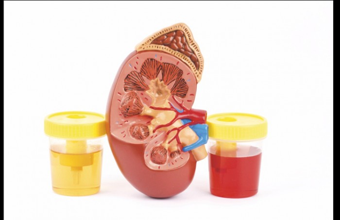
 Introduction:
Hematuria is a symptom of which the cause must be sought. The etiologies are numerous, and a rigorous diagnostic approach is necessary. The first stage of the assessment should confirm the diagnosis of haematuria, as false positives are frequent.Interrogation is a fundamental time of the etiological diagnosis.
The prevalence of microscopic hematuria in the general population varies from 0.19 to 16.1%. These large variations are related to the heterogeneity of the populations compared. In populations of men over the age of 60, Britton reports a prevalence of microscopic hematuria ranging from 13 to 20.1%.
Differential diagnosis:
The differential diagnosis must be made:
– with urethorrhagia: it is a flow of blood by the urethral meatus independently of urethral urination;
– with bleeding of genital origin in women (metrorrhagia, menorrhagia). If the woman is menstruating, the bleeding can contaminate the urine.
The analysis of the urine must therefore be remotely re-screened;
– with a red coloration of medicinal urine:
– several medicinal products may cause red or orange coloration of the urine, such as metronidazole, phenylindanedione, rifampicin, sulfasalazine, L-Dopa, ibuprofen, dantrone, phenolphthalein laxatives, polyvidone – iodine;
– certain foods may be involved: beets, blackberries …;
– with the presence of pigments in the urine (myoglobin, biliary pigments, melanuria, alcaptonuria, porphyrinuria);
– with a deliberate or artificial hematuria (Münchhausen syndrome). It is a diagnosis of elimination, which must be considered when no cause for hematuria is found. Chew reported the cases of nurses who harvested their blood by phlebotomy, then instill it into the bladder.
Positive diagnosis:
DIPSTICK:
It is a simple means of screening for microscopic haematuria.
Its sensitivity varies from 91 to 100%, and its specificity from 65 to 99%.
This test detects the presence of heme in the urine due to the peroxidic properties of the hemoglobin, which explains the false positives in case of hemoglobinuria or myoglobinuria. Lam showed that certain bacteria (Gram-negative bacilli, staphylococci) had peroxidase properties.
As a result, a urinary infection to these germs may be accompanied by false haematuria in the urine band.
The use of disposable plastic glasses for collecting urine is recommended as foot glasses cleaned with bleach or polyvidone iodine may induce false positives.
False negatives are very rare, and caused by acid pH. Demonstration of microscopic haematuria in the urine band should be confirmed by urinary sediment analysis. The etiological assessment should therefore not be started without confirmation of hematuria. The search for an associated urinary tract infection must be systematic. Indeed, 7% of microscopic haematuria are in fact urinary infections. It is then necessary to check the disappearance of the haematuria 6 weeks after the end of the antibiotic treatment.
DIRECT EXAMINATION OF URINARY SEDIMENT:
This analysis should be performed outside the period of menstruation, and at a distance (48 hours at least) from physical exercise or sexual intercourse. It is a semiquantitative method for the detection of microscopic haematuria and morphological analysis of red cells. Under standard conditions, 10 mL of urine should be centrifuged for 5 minutes at 2000 rpm. The urinary sediment is then analyzed using a phase contrast microscope. If the urine has been contaminated by the skin or vaginal mucosa, the sample should be redone.
Microscopic hematuria is defined as the presence of at least three red blood cells per field under a microscope, over two or three urinary sediment analyzes. In patients at high risk of malignant tumor (smoking or exposure to chemicals), evidence of microscopic haematuria on a urine sample is sufficient to justify a urological assessment.
The morphology of the erythrocytes can be analyzed at the same time. In the case of bleeding of glomerular origin, because of their passage through the glomerulus, the erythrocytes are deformed and of small size. Conversely, in the case of hematuria of extraglomerular origin, the erythrocytes have the same appearance and size as the peripheral blood. Shichiri, based on the fundamental assumption that glomerular hematuria was associated with small red blood cells, proposed using a hematology automaton to obtain curves representing the volumetric distribution of urinary erythrocytes to differentiate glomerular and extraglomerular hematuria.
The presence of hematic cylinders in the centrifugation pellet necessarily reflects the glomerular origin of haematuria.The hematic cylinders correspond to a stack of red blood cells on top of each other, the whole being maintained in this configuration by the protein of Tamm-Horsfall, which is secreted by the descending branches of the loops of Henle.
The cytobacteriological examination of the urine allows a quantitative analysis of the red cells in the urine. The diagnosis of microscopic haematuria is made when the number of red blood cells is greater than 5,000 / mL. The Addis account is less and less used because it is less precise and more random than the analysis of the urinary sediment.
Clinical evaluation:
INTERROGATORY:
Antecedents:
This time is fundamental for the diagnosis of hematuria.
It makes it possible to orient the prescription of complementary examinations:
– study of the characteristics of hematuria (initial, terminal, total, date of onset, number of episodes, associated signs);
– surgical, lithiasic, tumoral history;
– medical history (lupus erythematosus, coagulopathy, diabetes, arterial hypertension, sickle cell anemia);
– smoking;
– occupation (contact with aromatic amines, benzidine and dyes promoting bladder tumors);
– ongoing medicines, including anticoagulants and platelet antiaggregants. An underlying lesion should always be sought, the bleeding of which has been favored by the anticoagulant.
Avidor reports 25% of malignant tumors in macroscopic hematuria in patients on anticoagulants. Aspirin may be responsible for hemorrhagic cystitis by direct toxic action. Some drugs, such as cyclophosphamide, are also cytotoxic to urothelium;
– nephropathy (polycystic kidney disease) or familial uropathy;
Amylose;
– stays abroad, in particular in the Bilharzian endemic area;
– tuberculosis vaccination;
– history of pelvic radiation therapy;
– recent abdominal trauma.
Related functional signs:
The associated functional signs are:
– signs of bladder instability with pollakiuria, burns and urinary excretions;
– lumbago or renal colic. These pictures point to a pathology of the upper urinary tract;
– prostatism with dysuria or acute urinary retention leading to prostate disease.
PHYSICAL EXAMINATION:
It gives several indications:
– alteration of the general condition: recent weight loss and cachexia, asthenia in favor of tuberculosis or a neoplastic process;
– fever;
– arterial hypertension, edema, weight gain, purpura should direct to nephropathy;
– rectal examination seeks adenoma or prostate cancer;
– examination of the external genital organs: search for a left varicocele that may reveal kidney cancer, epididymitis in the context of urogenital tuberculosis;
– palpation of the lumbar fossa in search of a large kidney;
– disruption of painful lumbar fossa in cases of renal colic or acute pyelonephritis;
– gynecological examination: vaginal examination and speculum examination for cervical or cervical cancer invading the bladder.
Arguments for a nephrological origin of hematuria:
The nephrological origin of hematuria must be suspected before a proteinuria greater than 1 g / L, renal failure or hematic cylinders in the urine. Clinically, the presence of arterial hypertension, weight gain with edema or purpura lead to nephropathy. Several types of kidney damage may be responsible for hematuria. First, glomerulopathies may be part of a general systemic disease (lupus, vasculitis, septic conditions such as endocarditis or hepatitis) or isolated (eg, membranoproliferative glomerulonephritis, immunoglobulin A [IgA] nephropathy) .
Chronic interstitial nephropathies on the other hand (of infectious or medicinal origin) may also be accompanied by hematuria. Renal biopsy is the examination of choice to clarify the nature of the nephropathy involved.
Hematuria can sometimes be isolated. In this case, the urological and nephrological assessment including creatinine, proteinuria of 24 hours and search for hematic cylinders, is normal. Renal biopsies of these patients, when performed, frequently show structural abnormalities of the kidney, including IgA nephropathies. The interest of a renal biopsy in these patients is discussed, as it does not usually change the therapeutic management. The risk of progression to chronic renal failure is low. An annual follow-up is nevertheless necessary, in search of high blood pressure or proteinuria. Yamagata reported a series of 432 isolated microscopic hematuria followed for 6 years. He observed a complete disappearance of hematuria in 44.2% of cases, persistence in 43.7% of cases, urinary calculi in 1.4% of cases, and proteinuria without kidney failure in 10.6% of cases.
Indication of additional examinations:
IMAGING:
Abdominal X-rays without preparation and ultrasound of the urinary tract are the complementary examinations to ask for first aid for the etiological diagnosis of hematuria due to their simplicity and speed of access.
Pelvic computed tomography, intravenous urography (IVU) and renal nuclear magnetic resonance imaging (MRI) will generally be prescribed in a second step, depending on the results of the first-line examinations and the patient’s interrogation. The sensitivity of the various complementary tests prescribed must be well understood in terms of what the clinician is looking for. Ultrasound and computed tomography are the most sensitive tests for the detection of urinary stones, tumors and kidney infections. The UIV is historically the reference examination to carry out the etiological assessment of a haematuria. However, its sensitivity is insufficient for the diagnosis of parenchymal renal tumors. Ultrasound and computed tomography are the best examinations in this indication. For a kidney tumor confirmed by CT, the sensitivity of UIV for masses less than 2 cm, 2 to 3 cm, and more than 3 cm, was 21%, 52% and 85%, respectively. When the UIV reveals a kidney tumor syndrome, ultrasound and computed tomography (CT) must be performed to determine the size and solid or liquid character of the tumor. The computed tomography makes it possible to carry out an assessment of extension at the same time.
Ultrasound is increasingly used as a first line in the etiological assessment of hematuria because it is a non-invasive examination, easy to access, and gives very precise morphological information. Computed tomography is more often requested in addition to ultrasound. MRI does not increase sensitivity for the detection of kidney tumors compared to computed tomography. Renal MRI is requested in addition to ultrasound and computed tomography in case of diagnostic hesitations, or to specify the topography of a renal tumor syndrome.
Computed tomography is the best examination to detect calculations of the urinary tract, before ultrasound and abdominal X-rays without preparation. The sensitivity of the computed tomography for the detection of urinary calculi is 95%, compared with 52 to 59% for the UIV and 19% for ultrasound.
The uroscanner is an examination of choice to clarify the etiology of a renal colic. The density of the obstacle can be measured, making it possible to distinguish a calculation of a urothelial tumor from the ureter.
The sensitivity of UIV is better than ultrasound for the diagnosis of urinary tract tumor. Those of the uroscanner and the UIV have the same diagnostic sensitivity in this indication.
In short, in the presence of haematuria, complementary examinations should be guided by the suspected pathology.Abdominal X-rays without preparation and renal ultrasound are therefore the first morphological examinations required. For Jaffe, the initial assessment of microscopic hematuria includes cystoscopy, renal ultrasound, urinary cytology and cytobacteriological examination of the urine. The UIV is indicated according to him only in case of persistence of the microscopic haematuria at 3 months.
CYSTOSCOPY:
Cystoscopy is the best examination to look for a bladder tumor. It allows visualization of the bladder mucosa and ureteral orifices. This examination is carried out under local anesthesia in a patient with sterile urine. The rigid cystoscope or the flexible fibroscope can be used indifferently. The flexible fibroscope appears better to diagnose lesions located on the anterior lip of the bladder neck, thanks to retrovision.
The assessment of microscopic hematuria should include cystoscopy when patients are at high risk of developing bladder tumors. This population includes patients over the age of 40, and those under the age of 40 who are either smoking or in contact with chemicals toxic to the urothelium. Patients who do not meet these criteria are said to be at low risk. The risk of finding a bladder tumor in them is estimated at 1%. Cystoscopy should not therefore be proposed as a first-line treatment. However, if macroscopic hematuria or signs of bladder instability (in the absence of urinary tract infection) occur during follow-up, cystoscopy should be performed without delay. Cystoscopy is performed systematically in case of macroscopic haematuria (unless the origin of the hematuria is a renal tumor).
URINARY CYTOLOGY:
It is a cytological study of urinary smear, which looks for malignant urothelial cells. The sensitivity of urinary cytology varies depending on the grade of the tumor and the cytologist. It is 90% for grade 3 urothelial carcinoma and 10% for grade 1 carcinoma. The sensitivity is 80% for carcinomas in situ. Negative urinary cytology does not formally exclude a urothelial tumor. This test should be performed as a first-line treatment in patients at high risk of bladder tumor and in patients with irritative signs of lower urinary tract (pollakiuria, micturition burns), particularly for in situ carcinoma of the bladder. bladder. A positive urinary cytology confirms the need for cystoscopy.
ENDO-UROLOGICAL METHODS:
Percutaneous nephroscopy or soft ureteroscopy can be used to detect the origin of hematuria from the top of the device. Urothelial tumors can be resected and then analyzed. Sometimes papillary necrosis, papillary angioma or haemorrhagic papillitis can be recovered and may be coagulated.
RENAL ARTERIOGRAPHY:
This is not a first-line examination. Renal arteriography must be performed when a vascular origin is suspected of hematuria. Selective embolization may be performed in the case of arteriovenous fistula or false aneurysm.
Special cases of hematuria:
HEMATURIES OF THE CHILD:
Most hematuria of the child are of nephrological origin.
Urological causes are represented in the majority of cases by kidney stones, trauma and malformations of the urinary tract.
EFFORT HEMATURIES:
Macroscopic hematuria may appear on exertion. It may be associated with proteinuria. Examination of the urine at rest, in supine position, at a distance from any effort, is perfectly normal. A minimal urological assessment is indicated to ensure that there are no bleeding lesions, such as lithiasis.
The hematuria of isolated effort has no pathological character.
Patient follow-up:
In 8 to 10% of cases, no cause can explain the hematuria after the initial assessment. The risk of developing a malignant tumor in a patient with asymptomatic microscopic hematuria varies from 1 to 3%. The tumor develops most often within 3 years after the initial diagnosis of hematuria. Patients with asymptomatic microscopic haematuria with a negative initial etiological balance should nevertheless be monitored. Grossfeld proposes to review them 6, 12, 24 and 36 months after the diagnosis of hematuria, with urinary sediment analysis, cytobacteriological examination of the urine, urinary cytology and blood pressure measurement at each consultation. The purpose of this follow-up is to not overlook a bladder tumor. Complementary examinations performed during the initial assessment should be repeated in case of onset during macroscopic hematuria, abnormal urinary cytology or signs of bladder instability. If monitoring is normal for 3 years, monitoring can be stopped.
