 Introduction:
Proteinuria may be isolated, or may have kidney damage (primary or secondary). The criteria which will make it possible to classify a proteinuria are its transient or permanent character, its abundance, its composition and its selectivity, that is to say the presence of proteins smaller than albumin, renal function, association urinary sediment abnormalities (hematuria, leukocyturia),
Screening for proteinuria, which is based on the urine strip, requires the search for an etiology. Knowledge of the etiology will determine the modalities of monitoring, treatment and to specify to the patient the prognosis of the renal impairment.
Albumin is only one of the constituents of urinary proteins but its low-level presence, called microalbuminuria, has a prognostic value in diabetes and arterial hypertension. It is now well established that in glomerular lesions, the proteinuria level at the time of the discovery and subsequent development of nephropathy has a prognostic value.Finally, more and more experimental and clinical arguments suggest that reducing proteinuria is a therapeutic goal in its own right.
Biological diagnosis:
The methods of screening, quantification and analysis of the composition of proteinuria should be separated. Albumin is only one of the constituents of urinary proteins. Over the last decade, techniques have been developed for the determination of albumin in urine for concentrations of the order of micrograms per milliliter, called microalbuminuria.
SCREENING:
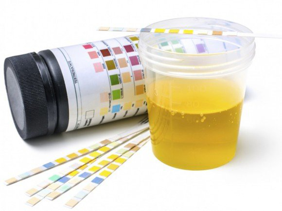
In terms of screening, two elements are essential to take into account: cost and ease of use. Screening for proteinuria involves the highly sensitive Albustix t or Multistix t urinary strips. Their principle is based on the binding of negatively charged proteins with the bromophenol blue dye, the coloring of which varies as a function of pH. The visual or automated result ranges from zero to four crosses. There are exceptional false negatives characterized by the exclusive presence of light (positively charged) chains of immunoglogulins.
The more frequent false positives are found if the urine is alkaline (pH > 7), too concentrated, or contaminated (urinary tract infection, menstruation).
Traces of albumin are detectable in the urine of healthy subjects but at levels not exceeding 20 mg / L.Microalbuminuria between 20 and 200 mg / L is usually not detected by the test strips, which have a sensitivity of 100 mg / L (trace) to 300 mg / L (+). “Three-drop t ” and “Albutest t ” are agglutination tests using human anti-albumin antibodies. They are used to detect concentrations
QUANTIFICATION:
Modalities of urinary collection:
Only the collection of 24-hour urine allows the quantification of proteinuria / albuminuria. Indeed, the dosages are carried out in mg / L and depend on the dilution of the urine (and thus the 24-hour diuresis). To ensure a good urinary collection, it is necessary to check that the creatinine urinary excretion, which depends on the muscular mass, is between 8 and 12 mmol / 24 h in the woman and between 10 and 16 mmol / 24 h in humans.
However, however simple it may be, the collection of 24-hour urine is rarely performed well. For a more pragmatic purpose, it is possible either to time the duration of urinary collection between two urinations (result expressed in μg / min) or to carry out the collection in the morning at the rising or during the consultation (result expressed then in mg / L). Precision and better reproducibility are optimized by the expression of the results relative to the creatinine concentration (expressed in mg of albumin / mmol of creatinine).
Methods of measurement:
Total proteinuria is usually assayed by a colorimetric method using pyrogallol red, with a sensitivity threshold close to 100 mg / L.
The quantification of microalbuminuria uses immunoassay methods, specifically measuring albumin, with sensitivity close to 1 mg / L and reproducibility greater than 95%. The radioimmunoassay method is considered the reference method. Equally sensitive is the assay performed by immunonephelometry or by Elisa (enzyme linked immunosorbent assay). Immunoturbidimetry, which is less costly for the laboratory, is equally specific but slightly less sensitive (5 mg / L). By definition, microalbuminuria is permanent. All microalbuminuria must therefore be confirmed twice over a period of 3 months.
The intra-individual variability of microalbuminuria is significant, ranging from 25% to 60%.
QUALITATIVE STUDY:
Physiology of the selectivity of glomerular permeability:
The glomerular filtration barrier (BFG) comprises three anatomical structures:
Fenestrated endothelium of the glomerular capillary;
The glomerular basal membrane (MBG) which is a network of glycoproteins whose mesh size is less than 4 nm;
– podocytes, the epithelial cells of the urinary chamber which rest on the MBG via pedicels connected together by a “slit membrane” which ensures a large part of the selectivity of the membrane.
This filtering barrier makes it possible to filter freely all the molecules whose radius is less than 2.6 nm. Beyond that, the passage of large molecules, especially proteins, is greatly hampered. Moreover, the negative charge of the glycoproteins of BFG prevents the passage of the majority of the proteins charged also negatively. The structures involved thus effectively oppose the filtration of plasma proteins which are macromolecules with a radius greater than 2.6 nm. Less than 1% of plasma albumin (40 g / L, radius close to 3.6 nm) crosses the BFG and is then found in the primary urine (5 mg / L). Then, before being excreted, 99% of the filtered albumin will be reabsorbed into the essentially proximal tubular structures. Albuminuria therefore only partially reflects a glomerular permeability disorder.
Physiologically, urinary proteins are composed of less than 20 mg / L of albumin, less than 50 mg / L of Tamm-Horsfall protein (mucoprotein synthesized in the broad ascending branch of Henle) and less than 20 mg / L of fragments immunoglobulin.
Electrophoresis and immunoelectrophoresis of urinary proteins:
Electrophoresis and immunoelectrophoresis make it possible to quantify the separated proteins as a function of their molecular weight and / or their charge, which are responsible for a differentiated mobility in polyacrylamide gel. An antiserum allows the precipitation of the complexes, then an automaton ensures the quantification. Depending on their size and weight, the glomerular and tubular proteinuria can be differentiated.
Proteinuria can come from four mechanisms:
– increased glomerular permeability;
– lack of tubular reabsorption;
– increase in tubular protein synthesis;
– filtration of small proteins in abnormal amounts.
An increase in glomerular permeability generates a proteinuria consisting of plasma proteins passing through the pores of the BFG. If the pores have a radius allowing only the molecules with a radius less than or equal to the albumin (Alb, 3.6 nm) to pass, the proteinuria will be said to be selective. If large proteins (such as immunoglobulins [Ig]) are found due to the existence of large pores or non-selective shunts, proteinuria will be said to be non-selective.In a first approximation, an Ig G / Alb ratio of less than 0.10 reflects the selectivity and directs towards glomerulonephritis with minimal glomerular lesions. Conversely, an Ig G / Alb ratio greater than 0.5 indicates a nonselective proteinuria found in the majority of glomerulonephritis but also in nephroangiosclerosis.
Physiologically, small proteins are freely filtered and then reabsorbed by the proximal tube. Tubular involvement results in the presence of proteins of low molecular weight in particular, beta-2 microglobulin and immunoglobulin light chains.Tubular proteinuria is usually close to 1 g / 24 h and rarely exceeds 2 g / 24h.
The filtration of small proteins in abnormal amounts in plasma is encountered mainly in myeloma. Monoclonal free (not associated with heavy chains) light chains (kappa or lambda) are produced in high amounts during myeloma. Small in size, they are freely filtered and then excreted in the urine, constituting Bence-Jones proteinuria.
Unfortunately, the light chains are not detected by the test strips and, in the absence of associated albuminuria (by reaching the renal filter), proteinuria may be false negative.
More rarely, this mechanism is involved in myoglobinuria postrhabdomyolysis and in hemoglobinuria intravascular posthemolysis (hemolytic anemias).
Finally, tubular secretion of proteins, particularly of Tamm-Horsfall proteins, is exceptionally recovered.
Measurement of the selectivity curve of the glomerular filtration barrier:
BFG lesions of multiple origins result in proteinuria. However, the amount of protein in the urine only partially reflects the extent of the glomerular permeability disorder. Indeed, the negative charge of the proteins prevents their filtration by the MBG, which is itself negatively charged. Moreover, the proteins, after having been filtered, are reabsorbed by the proximal tube to more than 90% for albumin, for example. Proteinuria is only an imperfect reflection of an increase in glomerular permeability to macromolecules.
The measurement of glomerular permeability to electrically neutral macromolecules (dextrans or ficolls) makes it possible to better quantify the disorder of glomerular permeability.
The reference method for studying glomerular permeability in humans is the measurement of the clearance of dextrans, which are exogenous, electrically neutral tracers that are neither reabsorbed nor secreted by the tubular structures. The filtered amount of dextrans is therefore exactly equal to the amount of dextrans excreted in the urine.The dextrans, contained for example in Rheomacrodex t , have rays between 2.6 nm and 6 nm. Their administration to humans makes it possible, after collection of plasma and urinary samples, to calculate clearances (ratio between the quantity excreted per unit of time and the plasma concentration) as a function of the size of the dextran.
For this purpose, in each sample obtained (plasma and urinary), the dextrans are separated according to their molecular weight by a size exclusion gel method. It is thus possible to trace the curve of sieving of the urine for each patient and to compare it with that of healthy subjects.
The interest of this technique is threefold:
– in pathophysiology, it allows to quantify the disorder of the glomerular permeability;
– in therapeutics, it allows to observe changes in glomerular permeability in response to different therapeutics;
– in research, it allows access to the parameters that govern the glomerular permeability to electrically neutral or negatively charged macromolecules.
RENAL TOXICITY OF PROTEINURIA:
Proteinuria translates into kidney damage, but conversely, it can also play a role in the progression of renal disease.The abnormal passage of proteins through BFG and mesangial tissue can induce glomerular involvement. Thus, it has been shown that transferrin, complement proteins and lipoproteins have a direct toxicity. This results in the production of growth factors, vasoactive products and, more generally, inflammatory proteins. This mechanism of own toxicity may partly explain the unfavorable prognostic character of proteinuria greater than 3 g / 24 h, regardless of etiology and blood pressure.
Reducing proteinuria by ACE inhibitors has even been shown to be beneficial at identical blood pressure.
Clinical significance:
ISOLATED BENIGN PROTEINURY:
Functional proteinuria:
It is very common and occurs during a fever, intense physical exercise, psychological stress, exposure to the cold. Its evolution is transient and the proteinuria disappears at 48 hours of the event. Renal prognosis is excellent.
In the pathophysiology, an increase in glomerular permeability and / or a decrease in tubular reabsorption was incriminated.
Transient Idiopathic Proteinuria:
This form is frequently encountered in children, adolescents and young adults. The screening is done in school medicine, military service or medical examination. There is no anomaly of the urinary sediment (neither hematuria nor leukocyturia). Before undertaking a more in-depth and inevitably more costly assessment, it is necessary to ensure that proteinuria is not disappearing from a distance.
Sometimes proteinuria is variable on several samples, which may be positive or negative. Renal biopsies performed resulted in lesions of fibrosis and / or increased cellularity. In these young individuals, proteinuria may disappear permanently, or persist, which should lead to monitoring of renal function.
Orthostatic proteinuria disappears, by definition, in the supine position. It is usually observed before 30 years and exceeds exceptionally 2g / L. Renal biopsies, carried out in the framework of studies, showed strictly normal glomeruli with very slight modifications in electron microscopy.
Orthostatic proteinuria decreases with age and often disappears into adulthood. Renal prognosis is excellent.
INSULATED PERSISTENT PROTEINURY:
The discovery of persistent proteinuria (independent of position and found on several samples) isolated (without urinary sediment anomaly or systemic pathology) corresponds to a disparate entity of nosology. In the rare longitudinal studies, the development rarely occurs towards renal insufficiency.
Renal biopsies performed routinely usually find normal kidneys, but sometimes have a tendency to mesangial hypercellularity and / or an increase in the mesangial matrix. As long as the proteinuria remains moderate ( < 1g / 24 h), renal biopsy can be avoided under the supervision of at least one year of renal function and proteinuria.
NON-NEPHROTIC PROTEINURY ASSOCIATED WITH RENAL ACHIEVEMENT:
Microalbuminuria:
The prevalence of microalbuminuria in the general population is in the order of 5 to 8%. In the course of the evolution of type I or II diabetes, microalbuminuria is an early glomerular disease. In the absence of diabetes, the presence of microalbuminuria has been found associated with other cardiovascular risk factors.
Tubular proteinuria:
Tubular proteinuria is usually close to 1 g / 24 h and rarely exceeds 2 g / 24h. Proteinuria consists mainly of beta-2 microglobulin and light chains. An amicrobial leucocyturia is often associated. Such tubular proteinuria is encountered in toxic nephropathies (heavy metals, analgesics), endemic nephropathy in the Balkans, chronic pyelonephritis, renal transplant rejection, but sometimes also in nephroangiosclerosis.
Glomerular proteinuria:
Proteinuria less than 3 g / 24 h may occur in most primary or secondary glomerulonephritis. It is usually non-selective.In France, it is found in order of frequency in the evolution of diabetic glomerulosclerosis, nephroangiosclerosis, Berger’s disease. Proteinuria greater than 1 g / 24 h is an independent prognostic factor (of blood pressure and etiology) with an evolution towards end-stage renal failure.
There is also glomerular proteinuria, rarely greater than 3 g / 24 h during pregnancy (characterizing preeclampsia), heart failure, stenosis of a renal artery (renin-dependent proteinuria).
NEPHROTIC PROTEINURY:
Nephrotic syndrome involves the association of edema, proteinuria greater than 3 g / 24 h and hypoalbumin less than 30 g / L.
Nephrotic proteinuria ( > 3 g / 24 h) can be found in most glomerular disorders. The selective or non-selective character of proteinuria, renal function, and the existence of an associated disease (lupus, diabetes, iatrogeny) as well as the age of the patient, heredity (Alport syndrome) and size of the kidneys can lead to the practice of a renal biopsy puncture (PBR) to establish histological diagnosis and therapeutics.
Independently of the etiology, a proteinuria greater than 3 g / 24h is a factor of poor prognosis for the evolution towards end-stage renal failure.
Selective Proteinuria:
Selective nephrotic proteinuria in pure nephrotic syndrome is only observed in minimal glomerular lesions with or without segmental and focal hyalinosis. In children between the ages of 1 and 10 years, the diagnosis is almost certain and corticosteroids will be carried out on the first intention without performing an ACB. In adults, an ACB will be performed to confirm the diagnosis before starting appropriate therapy.
Non-Selective Proteinuria:
The vast majority of primary or secondary glomerulonephritis may be accompanied by nonselective nephrotic proteinuria. The most frequently encountered glomerular lesions are diabetic glomerulosclerosis, extramembraneous glomerulonephritis, segmental and focal hyalinosis, poststreptococcal acute glomerulonephritis, renal amyloidosis, membranoproliferative glomerulonephritis and extracapillary glomerulonephritis. More rarely, nephrotic proteinuria may be present in IgA nephropathy (Berger disease, rheumatoid purpura), heavy metal poisoning (lead), HIV infection, nephroangiosclerosis, and stenosis of the renal artery.
Conclusion:
Proteinuria detected by the test strip ( > 200 mg / L) is a frequent reason for consultation in town medicine.Diagnostically, modest proteinuria ( < 1 g / 24 h) should be characterized before leading to heavy and costly explorations. First, the permanent or transient (fever, orthostatism) character of proteinuria should be confirmed. Renal function (calculated creatinine clearance) and urinary sediment abnormalities will finally determine the urgency of the diagnosis.
If proteinuria is permanent, electrophoresis and immunoelectrophoresis of the urinary proteins will reveal the tubular origin (tubular nephropathy) or glomerular (primitive or secondary glomerulonephritis) of the proteinuria as well as its selectivity. A lack of reabsorption by the proximal tube of the small freely filtered proteins (beta-2 microglobulin) results in tubular damage, whereas proteinuria rich in albumin reflects an increase in glomerular permeability.
A nephrotic proteinuria greater than 3 g / 24 h translates a glomerular disorder, which will usually lead to a PBR (apart from diabetes and nephrotic syndrome in children) to perform a histological diagnosis and adapt an appropriate therapeutic.
In terms of prognosis, microalbuminuria (albuminuria between 30 and 300 mg / 24 h) is a cardiovascular risk factor during diabetes or hypertension, indicating a systemic microvascular involvement with renal expression. A more abundant proteinuria will result in a more pejorative renal prognosis. Proteinuria reduction alone has been shown to be a therapeutic objective.
Finally, the more proteinuria is detected early, the better the therapeutic will be adapted and the better the renal prognosis.