DIAGNOSTIC:
 The pupillary examination is done in the light and in the dark, while the subject look away. We study compared the shape, square, pupil size, and responsiveness to light, one after the other.
If one or both pupils are aréactives to light, we search contraction during synkinesis accommodation-convergence.This is done by asking the subject to attach a small object that is approaching so that the eyes converge as possible to this item.
Sometimes you pursue this stimulation over a minute.
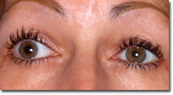
If anisocoria is discovered, it must date from old photos, especially if it is isolated.
Pupils normally reactive to light and are symmetrical normal pupils, even if they appear in miosis or mydriasis.
The causes of error are:
– Lack of consideration in the darkness that leads to a disregard Horner syndrome;
– Request by the examiner to fix a point close ( “look at me”) requesting the accommodation-convergence and leads through this miosis. We need the right patient fixed before him, in the distance;
– The necessarily sudden discovery of an abnormality sometimes old. The discreet anisocoria a subject with dark eyes, discovered during conjunctivitis can actually be very old.
Unlike frank about a blue-eyed anisocoria revealed by photophobia or discomfort in reading;
– The inadequate eyedrops may prevent correct approach later. False test corneal sensitivity some pharmacological tests.
ETIOLOGY:
Eye causes:
Some eye and not purely neurological diseases can cause pupillary abnormalities that are not in the foreground.
Acute glaucoma:
Acute glaucoma should be recognized by any doctor, and treated even in non-specialized areas, because it is an emergency in one or two hours near.
The table can be unilateral or bilateral. The patients presenting with orbital pain and / or headaches that may be accompanied by nausea.
The eye is often red. The existence of colored halos around lights is a strong diagnostic argument. In bidigitale palpation, the globe seems harder than that of the examiner or other control subject.
Unless an eye examination is possible within two hours, the treatment should be started before the transfer. It consists of a bulb Diamox® IV, and 4 measures glycérotone associated with beta-blocker eye drops, and only if the diagnosis is certain, pilocarpine eye drops every hour if the tolerance is good (to close your eyes 2 minutes after each instillation to reduce the systemic absorption).
Uveitis:
Uveitis can be affirmed by an examination at the slit lamp. This eye exam should be done urgently before any red eye, let alone if pupillary abnormalities (irregularities shift, asymmetry, abnormal light reflex).
For treatment, see Chapter Red Eye (uveitis).
Neuro-ophthalmologic causes:
Anisocoria:
After searching a toxic cause (atropine, nicotine, cocaine, etc.), especially in botany (belladonna, datura, etc.), we must remember three basic diagnoses (Fig. 1) by the severity, frequency, differential diagnoses :
– Horner syndrome (HBC) and in particular the dissection of the carotid artery;
– The compression of the third cranial nerve (III), in particular by an aneurysm;
– The pupil of ADIE, which bilateralization must seek primarily a diagnostic error (it is exceptional despite some erroneous figures of literature).

It combines reactive miosis, increased in the dark to a narrowing of the palpebral fissure due to a still moderate ptosis and an ascent of the lower eyelid (Müller muscle damage, innervated by the sympathetic).Horner’s syndrome:
This gives a false appearance of enophthalmos, but never true enophthalmos.
The photomoteurs reflexes are normal: the pupil dilates pathological evil but good contracts. The main differential diagnosis is anisocoria physiological and test to cocaine eye drops (Fig 2, see also figure in the color booklet.) May be required to resolve: its instillation into the eyes majorises anisocoria when syndrome CBH, and reduced in case of physiological anisocoria.
The sudden onset of CBH without further neuro-ophthalmological disorder, iatrogenic without context and without lymphadenopathy in the cervical palpation, is most often the result of a extracranial carotid dissection. If he joins in ipsilateral pain, it is always a carotid dissection, even if the subject relates his pain to a dental problem, sinus or another.
The risk is not breaking but as thromboembolism in carotid stenosis.
Compression III and ruptured aneurysm:
Any little or nonreactive mydriasis associated with diplopia is a priori a violation of the intrinsic and extrinsic compression and translated III of III. This shows the importance of the review of the pupils in the case of diplopia.
Any pain ipsilateral to impairment of the intrinsic and extrinsic III reflects an aneurysm cracking. The transfer neurosurgical environment must be immediate. MRI confirm the diagnosis.
Pupil ADIE:
Isolated acute or mydriasis associated with accommodative disorder (poor near vision) without diplopia or ptosis is usually a pupil of ADIE. But if the pupil nonreactive to light does not really contracted and toned way in accommodation-convergence, it is necessary to promptly differential diagnosis between pupil and paralysis of intrinsic ADIE III.
A fortiori in case of pain.
Bilateral mydriasis nonreactive:
Because of the urgency and public health implications, eliminate pharmacologic cause (see above), and recall biasbotulism whose best signs are the food environment and the achievement of several people, drought mouth, micturition disorders, digestive, neurological or that evoke a botulism.
Bilateral miosis unresponsive:
We must seek a pupillary contraction in accommodation-convergence: if present, this is:
– A rare bilateral pupil aged ADIE;
– Instead of p upilles Argyl Robertson.
Syphilis serology is systematic although most of the pupils are Argyl Robertson to diabetes.
Gunn pupil:
Someone with an optic nerve never leads to anisocoria. By cons, a lesion of the optic nerve (unilateral or asymmetric) can often be objectified by examining the pupils, by enlightening them alternately.
CONCLUSION:
The management of a pupillary anomaly follows almost mathematical logic and leaves no room for improvisation. It must be emphasized:
– The need to properly date pupillary defect and eliminate a pharmacological or toxic affected by specific issues before embarking on complex investigations;
– The urge to examine the pupils in the dark;
– Bad “umbrella” that is MRI if it is not oriented. In particular, whenever there is mydriasis reached by the Third while MRI is normal, that is that MRI ignores compression of III.
– A patient with a painful anisocoria may be dead (ruptured aneurysm) or hemiplegia (stroke secondary to carotid dissection) in the coming hours. Diagnosis and effective care possible to avoid it.
You must be logged in to post a comment.