 GROSSE LANGUAGE:
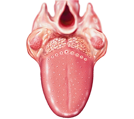
The language normally weighs 80 g in women and 97 g in man. The existence of a large tongue defines macroglossia.
His positive diagnosis seems immediate, but has two pitfalls: the differential diagnoses that are related macroglossies and protrusions lingual. The macroglossies on are represented by micrognathies, particularly in the context of trisomy 21 and Pierre Robin syndrome. The protrusions lingual meet if adenomegalies compressive, pharyngeal or mandibular deformity narrowness.
Among the true macroglossies (Box I) are distinguished by macroglossies increase of the lingual musculature of those due to infiltration by a substance or a tumor.
Box 1. Major causes of macroglossia
Macroglossies by increasing the lingual muscles
Hypothyroidism
acromegaly
Malformation syndromes: Beckwith-Wiedemann syndrome, Costello, Borjeson-Forssman-Lehmann, Tollner Manzke-Horst-Zimmerman-Laband, 48 XXYY syndrome, 14q partial deletion 14p partial duplication, triploidy, pseudomongolisme dysplasia panostotique fibrous, macrocéphalie- syndrome Member short deafness, coloboma, microphthalmia-heart-deafness syndrome
Warfarin embryopathy
beclomethasone
idiopathic macroglossia
Macroglossies infi ltratives or tumor
amyloidosis
sarcoidosis
Edema: angioneurotic, angioneurotic post-traumatic, post-surgical, post-radiotherapy
cysts
Infections: pyogenic, mycobacterial infections, syphilis, hydatid cyst, actinomycosis.
Cancers: squamous cell carcinoma, rhabdomyosarcoma, plasmacytoma, cancer thyroid remnants.
Phakomatoses: von Recklinghausen disease, Cowden syndrome
Lymphangioma, hemangioma
Melkersson-Rosenthal syndrome
storage diseases: disease Hurler, Hunter, pump, alphamannosidose, Aspartylglucosaminuria, gangliosidosis GM1
pemphigus vulgaris
Macroglossies by increasing the lingual muscles:
The macroglossies by increasing the lingual muscles may be related to the increase in myocyte size (hypertrophy) or number (hyperplasia).
Hypothyroidism:
The hypothyroid macroglossia occurs mostly the extremes of age and is linked to both lingual hypertrophy and myxedema.
The diagnosis is often simple (asthenia, nervousness, bradycardia, constipation, psychomotor retardation) and confirmed by TSH and possibly free T4.
Acromegaly:
Macroglossia of acromegaly is a lingual hypertrophy predominantly anterior, averaging 150 g.
The diagnosis is usually clinically evident and is confirmed by the assay of GH.
Malformation syndromes:
The most common is Beckwith-Wiedemann syndrome. Linked to an autosomal dominant mutation, it is usually sporadic, but sometimes inherited. It affects one newborn for 13,700 with a female predominance.
It is defined by a triad: macroglossia (usually unilateral hyperplasia), abnormal parietal closure (omphalocele, etc.), and body hemihypertrophy (may affect the internal organs: liver, kidney, pancreas). May be associated: hypoglycemia by hyperplasia of the islets of Langerhans, facial nevi, a bottom crease of the ear lobe and a microcépha binds. In over 10% of cases occur mainly at the expense visceral cancers of the kidneys, but also liver, adrenal glands, muscles and central nervous system.
Beclomethasone:
Rare cases of macroglossia have been described in premature infants treated over a month by this steroid.
Macroglossia gradually decreased to stop taking this medicine.
Idiopathic Macroglossia:
This is of course a diagnosis of exclusion.
This is a lingual unexplained muscle hypertrophy.
Or infiltrative tumor Macroglossies:
Amyloidosis:
This is the most common cause in adults. Macroglossia is mostly encountered during AL amyloidosis (20% in some series). It is rare in AA amyloidosis and exceptional in other types. It is typically indurated and sometimes tender to palpation.
The diagnosis is easy in a familiar context or evocative (nephropathy, neuropathy, heart disease). It is confirmed by biopsy of the salivary glands.
Sarcoidosis:
Sarcoidosis is a rare cause of macroglossia.
The diagnosis is often mentioned in the presence of mediastinal lymphadenopathy or interstitial lung lesions confirmed by biopsy of the salivary glands.
Edema:
Edema hereditary angioneurotic is linked to an autosomal dominant deficiency in C1 inhibitor.
It often presents as a circumscribed skin edema, transient, recurrent and non-pruritic. The respiratory disease is common and menacing mouth edema, tongue or larynx. Preventive treatment includes danazol and corticosteroids.Curative treatment is infusion of C1 inhibitor.
The angioedema is well known. Post-traumatic edema is of immediate diagnosis, in an epileptic context, accidental, surgical or radiotherapy. Installation is quick and involves a vital immediate risk for airway obstruction.
Cyst:
Mucoid or dermoid cysts lingual can present as localized macroglossies.
Infections:
Among infectious macroglossies, outside of pyogenic infections can be mentioned mycobacterial (especially atypical mycobacteria), tertiary syphilis reached (gums), hydatid cysts and actinomycosis.
These occur willingly after tooth extraction or trauma and appear as a painful indurated swelling.
Cancer:
Squamous cell carcinomas occur mainly in the éthylotabagique adult. Rhabdomyosarcoma are childhood tumors.
Their diagnosis is histological. Plasmacytomas lingual and cancers of thyroid remnants have been described.
Phacomatose:
Neurofibromas von Recklinghausen disease rarely affects the language.
They are then usually unilateral and often recurrent. The presence of other neurofibromas and café au lait spots diagnostic aid.
In Cowden syndrome, the skin of the face and extremities can wear papular lesions and the language can be the seat of hamartomas. These benign tumors can escalate.
Lymphangioma:
Lymphangiomas are the most common cause of children’s macroglossies. They are usually present at birth, but can develop at any age. They consist of dilated lymphatic vessels. When the lymphatic spaces are dilated cysts, they may form a particular variety: cystic bursitis. Macroglossia lymphangiomas can be localized or global.
It typically presents as a soft nodule back of the tongue.
Hemangioma:
Hemangiomas are the second cause of macroglossia. They are formed from dilated vascular spaces and reach all or part of the language.
Melkersson-Rosenthal syndrome:
It combines recurrent facial palsy, orofacial edema and asymmetric language Plicata possibly scrotal sometimes increased volume. His relationship with Crohn’s disease and sarcoidosis are uncertain.
Treatment consists of oral corticosteroids and / or general.
Enzymopathy:
They are characterized by substrate accumulation in the interstitium and some may give macroglossies. Among mucopolysaccharidoses, Hurler disease (autosomal recessive) disease and Hunter (X-linked recessive) respectively begin at 8 months and 3 years. Their manifestations are similar: hernias, facies gargoyle, multiple dysostoses, dwarfism, hepatosplenomegaly, cardiopulmonary impairment and mental retardation.
Among the glycogen storage diseases, acid maltase deficiency can reach the child or adult. It is autosomal recessive.In infants (Pompe disease), it is characterized by hypotonia and hypertrophic cardiomyopathy fatal within a year. In children and adults, it is as myopathy beginner belts but can win the respiratory muscles. Other storage diseases, even rarer, will not be developed here: alphamannosidose, Aspartylglucosaminuria, gangliosidosis GM1.
Pemphigus vulgaris:
This is an exceptional cause of macroglossia.
The context is bullous evocative. The diagnosis is confirmed by skin biopsy and antisubstance epidermal intercellular antibodies.
Complications:
The complications are many. Asphyxia is the major complication. Moreover, besides parental psychological shock in case of congenital macroglossia, dental deviations and prognathism, it should be mentioned lingual bites, difficulty swallowing and phonation.
Treatment:
The treatment is that of the case when it exists. If (risk of) complication, surgical lingual reduction is envisaged.
BLACK LANGUAGE:
Symptoms:
Patients consult for a black color more or less extensive language.
It starts on the midline back near the lingual V, and then extends forwardly and on the sides. The edges are not sharp but crumbled.
The hue ranges from light brown to anthracite black.
The language in this area takes a hairy appearance due to hyperkeratosis of the filiform papillae that extend abnormally.
Diagnostic:
The black tongue villous: in most cases this anomaly affects adults of all ages and evolves through time, over years, with a tendency to relapse. It appears no obvious cause. frequently we find a smoking or alcoolotabagique field.
If you realize a levy, often reveals the presence of Candida albicans and a varied bacterial flora saprophyte.
It is not sure however whether a mycological of affection but more likely a secondary colonization threadlike enlarged buds.
Antifungal treatments given in these cases are quite ineffective.
In a number of cases, we find these black languages of antibiotic treatment suites (often cyclins) that are due to candida overgrowth by breaking the balance of the local flora.
These include cases of radiotherapy treatment post-black tongues ENT often take a long time to fade after the end of treatment.
Treatment:
Post antibiotic black Languages:
It makes sense to give local antifungal therapy without having achieved sampling.
mouthwash is performed several times a day with products such as liquid Fungizone® the Daktarin® oral gel, gynecological Tablets Mycostatin® to suck.
Black hairy Languages:
For quite limited damage, make regular brushing and scraping of the tongue, badigeonnages with alkaline products such as baking soda or an alcoholic solution of 5% salicylic acid.
For large lesions, keratolytic solutions can be used tretinoin 0.05 to 0.2%: Apply the solution, leave for 2 to 3 minutes, then rinse well after brushing a soft, repeated twice daily.
GEOGRAPHICAL LANGUAGE:
Geographic tongue is known under different names: exfoliative glossitis migrans, peeling aberrant marginée areata.
Symptoms:
His appearance is quite evocative and rarely lets go wrong with other violations of the language. Is observed on the top and sides, a set of images in the form of spots, rings, festoons and circinations. Their appearance and their numbers vary rapidly over time. Thus the formed pattern can completely change within days. These lesions are bordered by a white border of one to two millimeters wide. The interior appears as abraded with a red hued degraded centripetally.
This is a particular type of keratinization phenomenon buds of the tongue that is more evenly but exfoliates selectively from a point centrifugally.
It is considered a variant of the physiological form. In most cases, this condition is painless. Very rarely it is complicated cracking and glossodynia.
Diagnostic:
For a long time its etiology has been ignored, at best we knew familial cases.
We spoke many triggers: foods, stress, infections, etc.
During his last years, some authors have considered this condition as a lingual localization of psoriasis. Others would have shown the existence of some familial cases to dominant transmission.
Differential diagnosis:
Despite its evocative appearance include such differential diagnosis: amylose, leukoplakia, lichen, syphilis.
Treatment:
The drug therapy is required. We must explain to patients that tested different treatments are very effective and very random. Furthermore, these lesions are evolving over time: appearing and disappearing quite quickly.