The subcutaneous emphysema corresponds to the appearance of gas in the subcutaneous tissue. It is either an air suffusion, spontaneous or during trauma, between an air cavity and the adjacent soft tissue (rupture of pleural bubble, tracheobronchial rupture, esophageal rupture) or a local production gas by anaerobic bacteria during infection (fasciitis and gas gangrene). More rarely the appearance of subcutaneous gas comes from a direct injection, often iatrogenic.
DIAGNOSTIC:
Clinically, it is a localized edema or soft tissue which can be diffused in the upper thoracic region and neck. Palpation allows to immediately recognize the diagnosis by finding a characteristic crackling snow under the fingers.
The diagnosis of subcutaneous emphysema retained, etiological research is directly related to the context: a debilitating disease or a torpid infection must immediately investigate and treat fasciitis, a reference to a trauma is considered its violence and its mechanism to search pneumothorax associated with rib fractures or tracheobronchial rupture (rare), the concept of local procedures (endoscopy, tracheal first, foreign body ingestion, dental) towards a local mechanism of injury.
Careful chest examination looking for signs related intrathoracic lesions (tympanisme auscultation and silence of a pneumothorax, mediastinal friction pneumomediastinum, etc.) and their complications (hemodynamic or circulatory failure).
ETIOLOGY:
Etiological research of a subcutaneous emphysema must be done urgently.
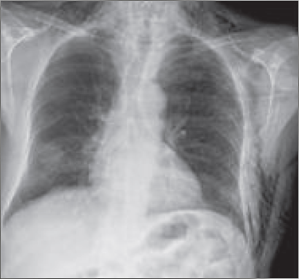
Depending on the diagnosis retained organizes patient monitoring. The diagnosis of thoracic subcutaneous emphysema is confirmed by a chest X-ray found that the presence of gas clarity in soft tissue to look carefully, especially at the clavicular hollow above (Fig. 1a and b).
It sometimes helps to find the cause: evocative pleural peel pneumothorax, rib fracture associated clear border molding the mediastinal silhouette, signing the presence of pneumomediastinum. CT scan allows to complete the study of pulmonary and mediastinal floor, and analyze possible tracheal and esophageal damage. Using a hospital structure depends on the intended aetiology and clinical impact. The “Quarantine” for 24 hours a spontaneous pneumomediastinum or secondary immediate cervicothoracic emphysema at a dental gesture can be discussed. The outpatient management may be considered when the environment, the absence of comorbidity and patient compliance permit.
Thoracic emphysema in a context traumatic:

Pneumothorax Posttraumatic:
Diagnostic:
The search for a chest emphysema is systematic in the management of all thoracic trauma especially during high kinetic accidents (public road accidents). The thoracic emphysema, however, can only appear secondarily after mask ventilation or mechanical. This sign most often the presence of a pneumothorax and / or pneumomediastinum. In high kinetic trauma, we must raise the possibility of a rupture of the tracheobronchial tree, which is rarely found (about 2.5% of cases) but that involves rapidly life-threatening.
Penetrating trauma (stab wound or fire) or external compression (strangulation) are rare in Europe.
They associate the lesions previously described more frequent vascular disease and skin sagging.
Emphysema can be very localized, next to a clinic or fracture site quickly spread to the neck and face. It is rare that the importance of emphysema requires a exsufflation own gesture. However, the pneumothorax may be complicated quickly and become suffocating by increased intrathoracic pressure (dyspnea, cyanosis).

Treatment:
The exsufflation needle to instantly reduce intrathoracic pressure and prevent the occurrence of respiratory and hemodynamic failure. It must be done without waiting either by anterior approach (2nd intercostal space, midclavicular line) or axillary (next to the 4th or 5th intercostal space, anterior axillary line). This simple and safe maneuver keeps the heart and breathing vital functions pending the drainage of the pneumothorax.
If pneumothorax blowing (stab wound or open trauma), it should ward off the plague of non-occlusive way to ensure the balance between intrathoracic pressure and atmospheric pressure.
Trauma of the tracheobronchial tree:
Diagnostic:
The injuries of the tracheobronchial tree are rare. They follow mostly an accident of the public highway (rib crushing or cervicothoracic, shearing after sudden deceleration, sudden vertical stretch by hyperextension of the neck), but can also complicate technical control air upper airway (endotracheal intubation, laser , esophageal tube-type Blackmore).Open injuries (stab or fire) are rare in Europe. These lesions are particularly sought before a cervical contusion, hemoptysis, dysphonia or a tension pneumothorax. Emphysema is predominantly cervical spine but often extends to chest or face with the intensity.
This results in three types of phenomena that could each lead a vital complication:
– Gaseous effusions in péritrachéo bronchial spaces in the mediastinum, the pericardium, in the pleural cavities;
– Bleeding into the tracheal lumen responsible for hemoptysis;
– Obstruction of the tracheal lumen responsible for respiratory distress.
The presence of a cervicothoracic emphysema in chest trauma or significant deceleration requires the rapid achievement of a CT scan that allows for the assessment of the lesions. Breach tracheobronchial endoscopy of the upper airway clarifies lesions, dictate taking initial therapeutic and should not be delayed. The mechanism of injury that the lung lesion is rarely isolated and a full review should be carried out, by not ignoring the brain lesions in 40% of cases.
Treatment:
The treatment of tracheobronchial lesions appealed urgently to selective intubation lesions and usually in reconstructive surgery (outside the membranous posterior longitudinal lesions, often iatrogenic).
Facial trauma:
Diagnostic:
Subcutaneous emphysema found in fractures of the air spaces of the face is common. It can sometimes spread in cervical and thoracic exceptionally causing pneumomediastinum.
Treatment:
The treatment is that the fracture of the facial structure.
Barotrauma:
Diagnostic:
Emphysema related barotrauma mainly observed during diving accidents.
The appearance of emphysema with the waning of a diving is related to the gas expansion nitrogen during ascent.Cervical extension is usual. This is decompression sickness which can be accompanied by a pneumothorax or pneumomediastinum.
He said against-the pursuit of underwater sports.
We can approach these barotrauma secondary alveolar ruptures with inhalation of a foreign body.
Treatment:
Treatment is based on HBO.
Iatrogenic:
Diagnostic:
Apart from recent surgery, emphysema can be found after tracheal intubation (tracheal or esophageal injury) but also after dental work. These were then mostly provided with a turbine.
The breaking of the lining with pressurized air injection causes the appearance of a cervical and thoracic edema, indolent. The extension may be towards the front. Complications from burglary subcutaneous are rare (optic nerve compression, secondary pneumothorax or pneumomediastinum), even if the original picture is sometimes impressive.
Emphysema related pneumomediastinum have been reported exceptionally in proctologies gestures after colonoscopy or laparoscopy.
Treatment:
Spontaneous reduction is the rule. However, it should check the time of onset of emphysema this with the care received (immediate to one hour). Antibiotic prophylaxis is common.
If times to onset is delayed relative to care, must fear a dental cellulite.
Infectious thoracic emphysema:
Fascitis Thoracic space is rare and usually secondary to infection complicating thoracotomy with sternal incision (sternotomy), chest wound or extension of an infection of the upper or cervical members on the trunk.
Bacterial cellulitis, necrotizing fasciitis with or without:
Bacterial cellulitis, necrotizing fasciitis with or without (DHBN-FN) is infectious achievement of subcutaneous tissues and includes the names of synergistic gangrene, gas gangrene (Clostridium perfringens), cellulitis, Fournier’s gangrene. It generally develops in adults over 50 years with associated comorbidities (diabetes, malignancy, immunosuppression and HIV, alcoholism). It is favored by the NSAID. This infection is often plurimicrobienne.
We gladly find the β-hemolytic group A streptococcus (S. pyogenes), with or without aerobic and anaerobic bacteria.
Diagnostic:
The front door is often a neglected chest wound, surgical intervention or the extension of a torpid DHBN-FN upper limb.
The infection develops in depth from an injury or sometimes minor trauma.
The first days, skin appearance is somewhat alarming to such ill-defined erythema. In 24 to 48 hours, the local aspect changes with an inflammatory appearance and tense, a purplish skin coloration. The appearance of skin bubbles containing a citrine or hemorrhagic fluid is indicative of the severity of the underlying process.
Purplish spots in geographic contours sometimes hypoesthesia next sign deep necrosis. General signs are then the foreground hyperthermia, prostration, hypotension or septic shock.
The secondary septic localizations are frequent. The local aspect worsens with extensive necrotic lesions and evolution is so often fatal. Thus early diagnosis is necessary. This unfortunately mainly based on the clinical appearance and the evocation of diagnosis. The presence of emphysema signs the presence of anaerobic and severity of the table.
Treatment:
Treatment relies on antibiotics surgical debridement of necrotic tissue and hyperbaric oxygen therapy.
Mediastinitis:
The outside mediastinitis cardiac surgery are rare, serious and high mortality.
The etiologies are mainly represented by the esophageal perforations (65% of cases) and oropharyngeal infections.The presentation is often severe at the outset due to the frequent diagnosis delay. The subcutaneous emphysema is the fact of polymicrobial flora usually with the presence of anaerobes.
Diagnostic:
The produced picture is that of a severe infectious process. The notion of an oropharyngeal infection or an endoscopic procedure in the days directs the diagnosis. The evocative functional symptoms are chest pain, cough and sputum, dyspnea and dysphagia. Quickly can appear a pasty and signs of sepsis leading to shock.
Again chest CT associated with esophageal opacification led to the diagnosis in 95% of cases.
Treatment:
Treatment is based on the broad antibiotic therapy directed against aéroanaéraobies germs, exploration and drainage of the mediastinal stage, and support care for organ failure.
Spontaneous thoracic emphysema:
Emphysema spontaneous onset or after a very modest effort without direct trauma usually corresponds to a pneumothorax or pneumomediastinum.
Pneumomediastinum:
Pneumomediastinum corresponds to the air outlet in the mediastinal cavity.
Spontaneous pneumomediastinum occurs after minimal exertion sports, singing, or after a closed glottis to stress such as childbirth, vomiting or defecation. It was also described in inhaled drug users or after taking ecstasy.
Diagnostic:
The clinical signs are poor and not very specific: chest discomfort remaining moderate, high dysphagia, dysphonia or dyspnea sometimes. The finding of a supraclavicular or cervical emphysema is found in nearly 90% of cases and remains the most constant physical sign.
Auscultation can find a sign of Hammam (creaking or grating noise synchronous mediastinal heartbeat, heard above the precordium and sometimes away from the thorax).
The diagnosis is not always easy because the chest radiograph allows only about 50% of cases to find the image characteristic of clear molding edging the mediastinum. CT scan allows however easily confirm the diagnosis.Complications from these spontaneous pneumomédiastins remain exceptional.
Monitoring of a few days to verify the ad integrum restitution. Diving is the only sport against-indicated after spontaneous pneumomediastinum.
Esophageal rupture:
Diagnostic:
The appearance of a pneumomediastinum during vomiting incentive to seek a spontaneous rupture of the esophagus (Boerhaave syndrome). Vomiting create the equivalent of a barotrauma rapid increase in intraluminal pressure on cricopharyngeal closure. Triad Mackler (chronologically: vomiting, and pain and subcutaneous emphysema) is evocative Boerhaave syndrome. This spontaneous rupture of the esophagus usually occurs after a heavy meal and effort vomiting. Often found in the history of these patients chronic alcoholism, obesity or an already pathological esophagus (reflux esophagitis, gastroesophageal reflux, ulcers, hiatal hernia, erosion of a tumor lesion). The break usually seat on the left side of the lower third of the esophagus.
But the majority of esophageal perforations are iatrogenic related to the development of endoluminal techniques (esophageal and tracheobronchial) and swallowed some drugs before bedtime insufficient liquid (cyclins, potassium chloride, bisphosphonates, NSAIDs) or secondary to the absorption wounding a foreign body.
Treatment:
The management of these perforations involves the combination of techniques déchoquage associated with antibiotic therapy and surgical rapid remedial action.
Thoracic emphysema in chronic bronchopathe:
Diagnostic:
Chronic bronchial diseases (COPD, emphysema, asthma) may decompensation during a pneumothorax or pneumomediastinum by breaking emphysema bubble or peripheral cells. The finding of emphysema of the sternal notch or supraclavicular fossa is evocative of a detachment of the mediastinal pleura.
Treatment:
The management depends on the tolerance of the effusion in relation to the underlying disease.
You must be logged in to post a comment.