 Definition-epidemiology:
The finger in mallet or mallet finger is a loss of continuity of the extensor apparatus on the back of the last phalanx, whose origin is traumatic.Segond, in 1880, made the first description of this deformation with a bony wrench. In 1887, Schoening described a subcutaneous rupture of the extensor without fracture. This lesion represents 2% of sports emergencies and is found in all sports with direct contact between the fingers and a ball. It occurs predominantly in men between 35 and 50 years of age, while 44% of women are more than 50 years old. The dominant hand seems a little more frequently reached than the non-dominant hand.
There are two types of lesions:
• “ tendinous mallets “ with subcutaneous rupture of the stent terminal strip in zone 1 of the Kleinert and Verdan classification;
• “ bone mallets “ , which can correspond either to the avulsion of the insertion of the stent on the base of the distal phalanx, or to a true fracture of the distal interphalangeal joint (IPD).
The bone mallets occur on average around 23 years and the tendinous mallets around 40-60 years. The frequency of the various lesions is variously appreciated in the published series. The long fingers are more frequently affected, especially those of the ulnar border of the hand.
Anatomical Considerations:
At the level of the long fingers, the extensor tendon divides with respect to the first phalanx (P1) into a central strip which is inserted on the base of the second phalange (P2) and into two lateral strips which meet to form the strips terminus of the stent after receiving the terminal fibers of the intrinsic muscles. The joining of the lateral strips takes place upstream of the distal insertion of the tendon on the third phalanx (P3). The stretch tendon stretch during IPD bending is 4mm. On the back of the proximal interphalangeal (IPP), the lateral strips of the extensor are maintained by the retinacular ligaments which are involved in the formation of the swan-neck deformation.
The lateral retinal ligament consists of the transverse and oblique retinal ligaments.
The transverse retinal ligaments are inserted on the lateral strips of the stent, after the joining of the fibers coming from the intrinsic. In palmar, they are attached to the A3 pulley and the flexor sheath in their proximal half, and to the palmar plate and capsule in their distal half. They cover the collateral ligaments from which they are readily separable. Their main function is to combat the dorsalization of the lateral strips when the finger is extended.
The oblique retinacular ligaments are inserted proximal on the lateral crest of P1 and terminate on the edge of the lateral strips of the extensor on the back of P3. Their function is to anticipate the flexion of P2 during the flexion of the interphalangeals to allow a gradual winding of the fingers. During flexion of P3, initiated by the deep flexor, they deflect the PPI, which has the effect of “ relax “ the IPD. The active flexion of the IPD is thus less in extension of the PPI (45 ◦on average) than in the full flexion of the PPI (86 ◦ on average).
The dorsal retinal ligament corresponds to the stretched structures between the lateral strips:
• the triangular ligament, which joins the two lateral strips on the back of P2 and whose fibers are mixed with those of the transverse retinal ligament;
• Arciform fibers, which pass above the joint, upstream of those of the triangular ligament, and which mix with the end fibers of the intrinsic tendons, mixing themselves with the central strip of the extensor.
Physiopathology:
The lesional mechanism of the mallet finger is a forced flexion of the IPD while the tendon extensor is contracted. It often occurs for minor trauma, which has raised the possibility of a familial predisposition and / or the existence of a weakly vascularized zone towards the distal insertion of the tendon. There also appears to be tendon embrittlement with age, which is more pronounced in women. Deformity can sometimes appear a few days after the trauma.
The fracture lesions are rather related to an axial impaction of P3 on P2 and are mainly seen in sports accidents. The tendon being adherent to the capsule, the tearing is often partial.
Diagnosis in the acute phase:
As a result of trauma to the distal end of the finger, the patient exhibits a characteristic flexural deformation of P3 which usually occurs immediately. This deformation is passively reducible, but the patient has lost all possibility of active extension of P3. In partial lesions, the deficit of extension is not very marked, but there is a decrease in net force in contracted extension. When the tear involves the tendon and the joint capsule, the flexion deformation is more straightforward.
In old lesions, the deformation becomes irreducible by retraction and adhesion of the oblique retinal ligament.
Face and profile X-rays are essential to detect avulsion or fracture of the base of P3. It is then necessary to specify the size of the bone fragment, its displacement and the possible existence of a palmar subluxation of the IPD. In case of fracture displaced, it is useful to carry out radiographs after the placement of the orthesis to appreciate the reduction of the bone fragment. In children, it may be a displaced epiphyseal detachment of P3.
Differential Diagnosis:
Few differential diagnoses are possible:
• Kirner deformity, a congenital anomaly of the distal phalanx of the long fingers. The age and the radiographic aspect of the deformation orientate towards the diagnosis;
• a rupture of the long tendon extensor of the thumb which must be evoked before a fall of P3 with pain and / or edema on the back of the wrist. Thumb-thumb testing is diagnostic;
• exostoses of the P2 neck, diagnosed on radiographs.
Spontaneous evolution:
In the absence of treatment, the evolution of the mallet finger takes place towards the proximal retraction of the extensor with the formation of a tendon callus which is too long. Anatomical studies have shown that a 1 mm elongation corresponds to a loss of extension of 25 ◦ , 36 ◦ for 2 mm, 49 ◦ for 3 mm and 63 ◦ for 4 mm. The proximal retraction of the lateral strips reinforces the action of extension of the median strip on the PPI and causes a hyperextension of the PPI in the lax subjects. The tension produced on the flexor deep increases the flexion of the IPD. Progressively, a deformation in swan-neck, first reducible, then fixed. Secondary retraction of the oblique retinal ligament prevents any correction.
Treatment of recent forms:
Orthopedic treatment:
It consists of an immobilization of the articulation IPD in straightness for a duration of six to eight weeks. It is the most frequently used treatment, but there is no consensus on its practical modalities. The splinting can be placed on the back of the finger or on the pulp, the position of the last phalanx is not precisely defined, the duration of immobilization varies according to the authors, as well as the temporary immobilisation of the l IPP articulation.
Type of splint:
Numerous publications have attempted to find benefits to the various commercial splints and / or performed by physiotherapists, whether dorsal or palmar. Katzman et al. have also described, with good results, the use of a removable “ flexible “ splint, taking two fingers in syndactyly and allowing only movements of 5 to 10 ◦ . However, no publication reaches a sufficient level of evidence to allow valid conclusions to be drawn and to recommend one type of splint rather than another.
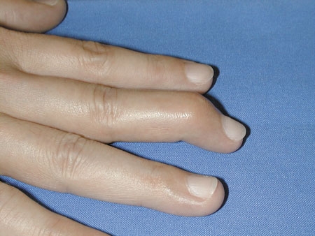
A good splint should be fairly sturdy, easy to use and comfortable. The main problem is tolerance. Mallet finger with palm subluxation of the IPD.
Surgical indication:
which often causes excessive sweating, skin irritation and discomfort in everyday life. In the Wehbe and Schneider series, 20% of patients prematurely dropped their splint. Variations have thus been described in order to avoid the support on the dorsal face of the IPD, which is often irritated. Most authors recommend regular washing of the hands and orthosis to avoid maceration. In order to wash the finger, the patient must place his hand flat on a table and, on the other, slide the splint to avoid any flexion of the IPD.
Position of immobilization:
Since PDI has an average passive hyperextension of 28 ◦ , some authors have advocated a hyperextension of P3 to promote healing. Although attractive, this method is not recommended because hyperextension of the finger increases the risk of cutaneous necrosis. In commercially available splints, it is possible to control the position in straightness by adding a piece of compress when the finger tends to keep a small flexion. In addition, in the bone finger fingers, the extension tends to displace the fracture and to increase the joint incongruence.
It is therefore essential to make X-rays to check the reduction of the fracture after insertion of the splint. The immobilization of PPI is not necessary except in patients with hyperlaxes or presenting immediately with a swan-neck deformation.
Duration of treatment:
The duration of immobilization varies according to the authors of six to eight weeks without interruption, followed by a period of night port for two to four weeks. The initial period may be extended in case of delay in initiation of treatment or persistent distortion in the removal of the splint.
When removing the splint, passive mobilizations of the IPD should not be carried out and any rehabilitation should be avoided which could lead to a strain of tendon elongation. Instead, it is necessary to practice reincarnation exercises in common gestures, while following him with a look to avoid trauma.
The resumption of the sport can be done under protection of a tight strapping of the last phalanx, but given the weight of the balloons and the speed of the impacts, this strapping is more a psychological support than a treatment.
Results:
Crawford, using his own criteria evaluated the results of 184 patients treated with splint.
These were generally comparable in patients treated early or secondary, but the poor results were slightly more frequent in the case of bone mallets.
Mikic and Helal, using the criteria of Stark et al. , found 40% good results; 53.3% of patients improved and 6.6% of patients unchanged. Of these, 80% had a complete flexion of IPD, but 20% had a flexion deficit after they had worn the splint longer. Forty percent of the patients had a complete extension, 40% had a deficit of less than
10 ◦ and 20% had a deficit of 10 to 20 ◦ . Four mild relapses occurred long after wearing the splint.
In the Niechajev series, subjective results were excellent in 82% of patients. Among them, 9% complained of minor disorders and 9% of major disorders.
The extension deficit was:
• nil in 70% of cases;
• less than 10% in 12% of cases;
• between 10 and 20 ◦ in 12% of cases;
• greater than 20 ◦ in 6.5% of cases.
A flexion deficit was noted in 11% of cases, but less than 10% of the time.
In the bone mallets, Lee found a decrease in mobility in 46% of cases, with loss of extension in 32% of cases.Fourteen percent of the patients complained of mild pain, particularly cold, but none had pseudarthrosis or joint pain despite joint deformity. Wehbe and Schneider, and more recently Kalainov et al., Have reported good results after orthopedic treatment of fractures involving more than one-third of the joint surface and associated with palmar subluxation. The final extension deficit was 9 ° æ11 ◦ and the flexion of 59 ° æ14 ◦ . However, patients were discerned from the dorsal prominence of the IPD, radiographs showed an interesting subluxation of 30% of the joint surface in the initially subluxed forms and 90% of the patients had signs of early osteoarthritis at only 3, 5 years of average decline. These figures are close to the 65% of osteoarthritis reported by Wehbe and Schneider about 17 bone mallet fingers that are more than 1/3 of the joint surface and are treated conservatively or surgically. The quality of the articular remodeling has also been stressed by many authors and justifies, for them, a conservative attitude. Weber and Segmüller recommend that bone mallets be used only in palmar subluxation, irrespective of the size and displacement of the bone fragment. In fact, Husain et al. have shown that palmar dislocation of IPD should be discounted when the bone fragment exceeds 50% of the articular surface.
There are two main factors of failure in orthopedic treatment, with early recurrence as soon as the splint is removed: on the one hand, a delay in the management with the interposition of a fibrous callus preventing the reduction and, on the other hand, intra-articular dislocation of the proximal fragment in the bone mallets. However, do not rush to talk about failure because there is a spontaneous improvement of 10 to 15 ◦ within six months of treatment. For Pulvertaft, orthopedic treatment gives 60% good immediate results while 20% of patients improve sufficiently thereafter.
Thus, in the series ofWarren et al., Of the 30 patients who refused surgery after failure of the orthopedic treatment, 13 (43%) spontaneously improved within the next six months.
Surgical treatment:
As early as 1930, the failures of the orthopedic treatment led the surgeons to operate the fingers in a mallet. However, Robb’s evaluation of the results in 1959 showed that early operative treatment was neither necessary nor desirable.This view is currently shared by the majority of authors who rarely propose surgery because of its iatrogenesis.
Percutaneous techniques:
The temporary axial stitching of the IPD, used for bone mallets without palmar subluxation, gives good and excellent results according to Hofmeister et al. However, the spindle tends to pass into the fracture line preventing reduction of the fragment. This is why some authors have proposed a direct broaching of the bone fragment associated with axial pinning of the IPD.
The technique of Ishiguro et al., Proposed in 1988, is currently one of the most used for bone mallets. In high IPD bending, a first spindle (9 or 10/10) is inserted at 45 ◦ in the IPD spacing, 1 to 2mm behind the fragment and is fixed in the P2.
The principle is based on the periosteal continuity which allows the reduction of the fragment during the flexion of the finger. The straightness of the finger then brings the bone fragment into abutment against the spindle, which automatically reduces the fracture. A second pin then sets the assembly at 30-40 ◦ bending. For lesions seen late, Ishiguro et al. propose to refresh the focus of fracture in percutaneous with the point of a needle. Several studies have analyzed the results of this technique according to the Crawford criteria. Pegoli et al., In a series of 65 patients, reported 46% excellent results, 32% had good results, 20% average results and 2% had poor results. Darder-Prats et al. report 18 excellent and three good results on 22 cases. Finally, Hofmeister et al., Of 24 patients, reported 74 weeks of follow-up, 38% had excellent results, 54% had good results and 8% had average results. Three superficial infections and two displacements of less than 2 mm were noted in this series.
Open-air techniques:
The approach is done on the back of the IPD by a skin incision in Z, S or H. The dorsal skin is fragile and must be treated with attention because the postoperative cutaneous necroses are frequent. Care must also be taken not to damage the nail matrix which begins distally just after the insertion of the extensor tendon.
The tendinous mallets are treated by direct suture with 6/0 absorbable thread if the distal tendon portion is large enough to accept a suture. Otherwise, a pull-out pull suture should be made with a push button on the pulp, which is generally associated with axial pinning of the IPD. Mini-anchors can also be used, although they are often voluminous for the distal phalanx.
The bone mallets with a fragment of small size, not accessible to osteosynthesis, are treated in the same way as the tendinous mallets. The bone mallets with a large fragment can be treated by different techniques: pull-out, single or double pinouts, mini-anchors, mini-screws, or even bracing.
Results:
Auchincloss compared in a randomized study the treatment with Stack splint and distal interphalangeal broaching without showing significant difference. Patients were very satisfied in 89% of cases and satisfied in 11% of cases.Ninety percent of the patients treated with splint were very satisfied, 5% were satisfied and 5% were dissatisfied. Other studies have reached the same conclusions, for both the bone and tendon mallets. Joint fractures give similar results regardless of the type of treatment, with the exception of sensitivity to cold which is increased after surgery.Radiological changes (beak or joint incongruence) are identical in the two treatment groups and occur in one third of the cases.
The study by Stern and Kastrups compared the results of 84 orthopedic treatments and 45 surgical treatments.According to the criteria of Stark et al., The result was excellent in 57.8% of operated patients and in 50% of nonoperated patients. Functional improvement was observed in 36.8% of operated patients and in 31.8% of patients without surgery. Treatment was unsuccessful in 5.2% of operated patients and in 18.1% of patients without surgery.The splints gave 40.4% of complications, with the majority of dorsal cutaneous ulcerations and nail-like deformities.The surgery gave 53% of complications of which the most frequent were infection, nail dystrophy and incongruence articular. Results in loss of extension were similar or identical between the splint and the surgery, but there was a clear difference in the loss of flexion in disfavour of the surgery.
In a series of 160 patients, Wehbe and Schneider reported 33% of complications after surgery versus 9% after orthopedic treatment. King et al. reported 41% complications on 59 operative bone mallets, including 14 partial cutaneous necrosis, eight recurrences, four pin infections, four nail dystrophies and two osteitis. All the fractures, however, have consolidated.
Overall, the functional results of the surgery are generally comparable to those of orthopedic treatment, but at the cost of more frequent complications, dominated by skin problems. If this iatrogenesis is unacceptable to some, the indications for surgical treatment should be applied sparingly.
Indications in recent forms:
The majority of mallet fingers are orthopedic. First-line surgery should be reserved:
• large fractures (greater than 1/3 of the articular surface) displaced and not reducible by the splint;
• non-reducible palmar subluxations;
• displaced epiphyseal detachments in children;
• open fractures.
However, these findings remain controversial in the literature, as shown by the recent work of Kalainov et al. which finds no difference between orthopedic and surgical treatment for bone mallets with or without subluxation.
Treatment of old forms:
An old or chronic mallet finger corresponds to a lesion that is more than four weeks old and not immobilized. This includes simple reducible mallets seen beyond the first month, as well as mallets dating back several months or years that are more complex to handle. Different factors must be taken into account:
• the mobility of IPD because a conservative surgery is considered only on a flexible articulation;
• the state of the cartilaginous surfaces of IPD which degrade rapidly by limiting the therapeutic possibilities;
• the existence of a swan-neck deformation which may impose a “ rebalancing “ of the extensor apparatus.
Orthopedic treatment:
Garberman et al. have retrospectively studied the results of orthopedic treatment initiated early (before the second week) to those treated after four weeks. He found no significant difference between the two groups, regardless of the type of splint, and whether or not there is a small bone fragment (always less than one third of the articular surface).Facca et al. recommend that they do not hesitate to begin orthopedic treatment beyond six weeks, provided that the number of days between the accident and the placement of the orthosis is added to the eight weeks of immobilization.Similarly, Obert advises to attempt orthotic treatment for any soft chronic mallet finger less than four months old. Good results have indeed been reported for orthopedic treatments started at eight or even 18 weeks.
Surgical treatment:
Tenodermodesis:
Among the various techniques for shortening the tendonial calf, the tenodermodesis of Brooks and Graner, developed in France by Iselin et al., Remains the most used. It consists of correcting the extension deficit by a monobloc elliptic excision of the skin and distended tendon callus, 3 mm wide at the back of the IPD, followed by a mass suture, edge to edge, to the nonabsorbable wire . Pinout of the extended IPD is typically associated for four to five weeks, but immobilization by splint is preferred by some because of its lesser iatrogenesis. In their series, Iselin et al. obtained 85% satisfactory results with an average flexion of 62 ◦ and an average extension deficit of 11 ◦ . Kon and Bloem obtained similar results with an average extension deficit of 5 ◦ and a 60 flex flexion on a series of 27 cases. Levante et al. emphasize however that the tendon shortening is difficult to adjust and that there is a risk of stiffness in extension if the shortening exceeds 3mm.
In the case of a swan-neck deformation with moderate hyperextension of the PPI, the tenodermodesis may be associated with a capsulorrhaphy of the palmar plate of the PPI by a Bruner-type approach. This involves performing excision in the orange zone of the palmar plate on 2 to 3 mm, followed by a tensioning by suture edge to edge, in order to obtain a 15 flex flexion of the PPI.
Tenotomy of the central strip:
Described by Fowler, this process allows the treatment of the swan-neck deformation by resection of the central strip on the back of P2, which has the effect of relaxing the stent apparatus. An immobilization of the IPP in extension, leaving the IPD free, is necessary during 15 days.
On a 20-case series, Grunberg and Reagan obtained a constant correction of gooseneck, but also four deficits of PPI extension and one case of complete loss of mobility.
Chair reported six excellent results, with complete correction of the deformation, and two partial results on a series of eight cases, without observing any complication. Finally, Lucas reported three total corrections, six good results and two failures on 11 cases. It is therefore a simple and effective technique but which, however, can cause deformation in the buttonhole of difficult treatment.
Reconstruction of oblique retinal ligament:
In 1978, Thompson et al. have described a tendon graft of the oblique retinacular ligament called the oblique retinacular ligament spiral (SORL) for the treatment of swan-neck deformation. This technique has been taken up by different authors with minor modifications. It performs an active tenodetic effect with simultaneous correction of the flexion of the IPD and the extension of the IPP, using the tendon of the small palmar. Two routes are required to pass the transplant to the dorsal surface of the IPD and to the palmar surface of the PPI. The distal attachment is made using a pull-out or a mini-anchor. Proximal fixation usually requires an intraosseous tunnel in the first phalanx, but Kleinmann and Petersen have described fixation to the flexor sheath.
The adjustment of the tension is delicate and must be done at 0 ◦ of flexion-extension of the IPD for 20 ◦ of flexion of the IPP.
In the suites, the graft is protected by a splint for three to six weeks.
In their article princeps, Thompson et al. reported 10 cases with only two moderate PPI extension deficits (10 and 15 ◦) and three deficits in the PDI (twice 10 ◦ and one </s> 15 ◦ ). They found a case of buttonhole, linked to excessive tension of the graft, which required an elongation of the transplant. Girot et al., In their series of 23 cases of SORL, obtained 45% complete correction of gooseneck, with 95% correction at the PPI level and 72% at the IPD level.However, they deplored 14% of buttonholes, 12% of axial or lateral deformations and 6% of recurrences. Kleinmann and Petersen also obtained three cases of buttonhole on 12 patients, two of which necessitated a surgical recovery.
Indications in the old forms:
Orthopedic treatment may be attempted until the fourth post-traumatic month whether or not the patient has been treated at the initial stage. The orthosis must be carried as much time as acute (eight weeks permanently, then two weeks at night) and according to the same principles.
Surgical treatment is necessary in case of irreducible flessum, inveterate dislocation of IPD or chronic mallets older than four months when there is a functional discomfort (usually for an IPD flessum greater than 30 ◦ ). Conservative surgery is only considered on soft fingers or softened by rehabilitation. An isolated chronic mallet is classically referred to as the Brooks and Graner tenodermodesis. In the case of swan-neck deformities, SORL, tenodermodesis with IPP capsulorography or Fowler tenotomy can be proposed according to the schools. On the other hand, if there is an osteoarthritis or an irreducible flessum despite the rehabilitation, the arthrodesis of the IPD is the only option conceivable.
Conclusion:
The most recent mallet fingers are most often splinted. It must be worn continuously for eight weeks and changed weekly without ever bending a finger. Nocturnal port until the tenth week and absence of reeducation limit recurrences by lengthening the tendinous callus. There is a consensus for emergency surgical treatment of displaced fractures exceeding one third of the articular surface and palmar subluxations not reducible by the splint, as well as for displaced epiphyseal detachments in children. In the chronic phase, orthopedic treatment may be offered until the fourth posttraumatic month. Beyond that, surgery is most often required with different techniques that are a function of the mobility of the IPD, the existence of an osteoarthritis and / or a deformation in swan-neck.