 Trapped popliteal artery syndrome, with its non-specific and fluctuating symptoms, is not always easy to diagnose. It is nevertheless an arterial disease whose evolution exposes young and sporty subjects to severe vascular complications, justifying, most often, a surgical treatment.
This is why it is important to know when to look for this condition and how to prescribe complementary examinations that require particularly specific and specific protocols.
A pathology still unknown:
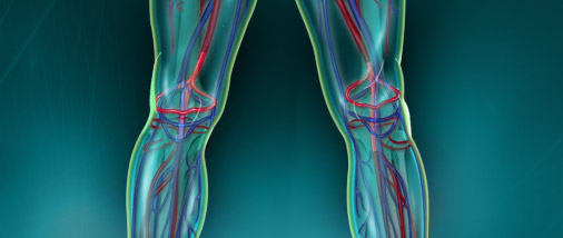
Trapped popliteal artery syndrome (SAPP) corresponds to the compression of the popliteal artery by the neighboring musculo-aponeurotic structures, during the movements of extension and dorsiflexion of the foot.
In the face of chronic leg pain in a young and athletic subject, one must know how to look for this syndrome.
Its early diagnosis is essential to avoid the evolution of the simple compression stage towards a chronic obliteration table with lesions of the arterial intima (fibrous stenosis, post-stenotic aneurysm) or acute arterial obliteration (by thrombosis of the l popliteal artery or distal artery embolism).
However, it is a pathology still unknown, whereas Stuart had described in 1879 an anomaly of the path of the popliteal artery on an inferior limb amputated for gangrene. In 1959, the first clinical case with surgical treatment was published and the term “popliteal artery entrapment syndrome” was proposed by Love and Welham in 1965 (2-3). Since then, the publications have multiplied and the nosological classifications succeeded one another.
In practice, there are three etiological groups:
* abnormal insertion of the proximal end of the gastrocnemius muscle on the femoral condyle (too high, too short or too external); it may also be a supernumerary muscle beam or fibrous tape trapping the artery (about 50% of cases);
* arterial path abnormalities: the popliteal artery is deflected, usually passing in and out of the gastrocnemius muscle, resulting in compression in muscle contractions (about 30-40% of cases);
* compressions of functional origin: arterial compression is due to the hypertrophy of the muscles of the popliteal hollow, following an important sporting training.
These abnormalities can be isolated or combined. They may also be associated with a popliteal vein trap.
Clinical features:
Population at risk:
The prevalence of SAPP in the general population is poorly known (a few studies estimate it between 0.2 and 3.5% of adult subjects).
However, it is known that this syndrome has a clear male predominance (according to the publications, between 75 and 93% of the patients are men). It mainly affects young and sporting subjects (more than 60% are under 30).Impairment is bilateral in about 30% of cases. The sports most often incriminated are those favoring the hypertrophy of gastrocnemius muscles: cycling, dancing, swimming, walking, rugby, football …
In the particular case of functional SAPP, the pathology appears to manifest itself earlier and affect women more frequently.
Functional signs:
The pathology is initially manifested by an intermittent claudication of the leg, uni- or bilateral, progressive aggravation, type of pain or calf cramp. This claudication is regularly paradoxical, more marked in walking than in running.
Its appearance may fluctuate from one day to another. The pain can appear by practicing certain sports and not others.
In more advanced cases, we can find a classic picture of intermittent claudication on exertion, or even signs of acute or subacute arterial ischaemia in the case of popliteal artery thrombosis or distal arterial embolism.
In case of associated venous trap, the patient may report leg or ankle edema, prolonged standing or exertion.
Physical examination:
It is usually normal or rough.
It must be bilateral and involve sensitization maneuvers: the active plantar flexion contraindicated and the passive dorsiflexion of the foot, the knee being in hyperextension. During these maneuvers, there is sometimes a diminution or abolition of the pulse at the posterior and pedic tibial level.
During palpation of the popliteal cavity, an arterial aneurysm can be discovered.
The absence of a popliteal, posterior or pedic tibial pulse in a young subject without atheromatous risk factor should evoke an SAPP.
Vascular echo-Doppler with dynamic tests:
This is the first-line examination.
It is non-invasive, inexpensive and reproducible, but operator-dependent.
First step: an examination at rest
First, the Doppler echo should study, at rest, systematically, the arterial and venous network of the two lower limbs.Indeed, even if they are sports subjects, often young, it is necessary to check the absence of arteriopathy obliterans of the lower limbs, which can be favored by smoking, genetic factors, diabetes or dyslipidemia unknown . This examination must be all the more careful since an arterial stenosis of less than 50% in reduction of surface does not modify the Doppler signal at rest, but can become symptomatic during an important effort. The absence of severe venous insufficiency, which may cause painful symptomatology, is also checked.
At the initial stage of simple arterial compression, resting of a patient with SAPP is usually normal. Sometimes an abnormal pathway of the popliteal artery or an accessory bundle of the gastrocnemius muscle passing between the artery and the vein is visualized.
At a more advanced stage, fibrous stenosis can be visualized, secondary to repeated arterial compressions.
This lesion may be associated with a post-stenotic aneurysm and endo-luminal clots. Populate artery thrombosis (or subpopliteal arteries due to distal arterial emboli) may also be observed.
Second step: sensitization through dynamic tests
Positional maneuvers aim to put the musculo-aponeurotic elements responsible for the popliteal trap in tension and trigger arterial compression.
In practice, these dynamic tests can be performed in a cooperating patient, in supine or ventral decubitus.
The help of a second examiner is strongly recommended to immobilize the lower limb examined in extension by placing a hand on the knee while the other can either mobilize the foot in passive dorsiflexion or resist a progressive active plantar flexion .
Meanwhile, the dopplerist analyzes blood flow at the popliteal artery if the patient is in a ventral decubitus or at the anterior tibial artery if the patient is supine. This study of the arterial flow should be as accurate as possible, in order to follow the hemodynamic variations while the foot changes its position. Examination is facilitated by the combined use of two-dimensional ultrasound with color and pulsed Doppler. An SAPP is suspected before a complete cessation of the flow during the passive dorsiflexion of the foot or during its active and non-maximal active plantar flexion.
When positional maneuvers are stopped, the suspicion of SAPP can be confirmed by a reactional hyperaemia, which is reflected on the Doppler signal by the appearance of a diastolic flow.
A dynamic load test can also be performed by asking the standing patient to tiptoe. If the Doppler echo shows compression of the popliteal artery with interruption of arterial flows downstream, a popliteal trap should be evoked.
In order to carry out this examination in the right conditions, it is necessary to plan a sufficient time slot and not to hesitate to repeat the maneuvers while being assisted by a second examiner.
When these dynamic tests are negative, the diagnosis of SAPP can be ruled out. In case of positivity, postural maneuvers should be interpreted with caution, because they are positive in 20% of normal subjects without a symptomatic popliteal trap. An abnormality during maneuvers must necessarily lead to further examination before making the diagnosis of SAPP.
Additional tests:
These are the additional examinations necessary to confirm the diagnosis.
Arteriography of the lower limbs:
This examination is carried out bilaterally after puncture of the femoral artery, with incidences of face and profile, the patient being in indifferent position and then carrying out the dynamic tests. It allows to detect a possible contralateral asymptomatic attack and to eliminate other arterial pathologies such as atherosclerosis.
In the case of SAPP, there is an anomaly of the path of the popliteal artery, with a medial deviation. It is also possible to demonstrate a stenosis of the popliteal artery, often described in an hourglass, the two extremities of which do not strictly have the same axis on the incidence of the face, due to arterial angulation. The examination may show any associated lesions, such as post-stenotic aneurysm or arterial thrombosis.
Currently, this technique is mainly considered when a revascularization is necessary and that one must visualize the upstream artery and the downstream bed. Arteriography has its status as a reference examination supplanted by more recent, less invasive examinations, also allowing a study of muscular abnormalities: computed tomography and, above all, magnetic resonance imaging.
CT and MRI:
The CT scanner can be used to diagnose anatomical abnormalities and show signs of arterial compression.
It must be sensitized by the same dynamic maneuvers as those practiced in supine position during Doppler echo.
MRI becomes the reference examination to confirm the diagnosis of SAPP.
It makes it possible to specify the path of the popliteal artery and its relations with neighboring musculo-aponeurotic structures, often denoting the causal anomaly. Thanks to the angio-MRI, it is possible to study the condition of the arterial tree in the undifferentiated position. The examination can then be completed with the dynamic maneuvers. In practice, with the intervention of an examiner in the MRI tunnel difficult, the patient is asked to hold a strap with a loop around the end of the strap. By pulling on the strap, it can carry out only the passive dorsiflexion and the active plantar flexion, against resistance, of the foot.
We can then observe an interruption of the popliteal flow, more or less complete, in sagittal sections.
Once the diagnosis is confirmed, the evolutionary potential of the SAPP requires surgical treatment, which includes the section of the musculo-tendinous elements that bridle the artery and, in the case of fixed arterial lesions, a vascular repair time. The earlier the diagnosis, the simpler the surgical procedure and the better the results at a distance (96% good results at the stage of simple compression) (11).
In conclusion:
Trapped popliteal artery syndrome is an often unrecognized condition, which has to be systematically investigated in a young, athletic subject with chronic leg pain with no uncovered cause. If the Doppler echo with dynamic tests is negative, the suspicion of popliteal trap can be ruled out. Otherwise, the diagnosis must be confirmed with the help of MRI, possibly scanning and arteriography, by performing dynamic maneuvers. Early and safe diagnosis requires multidisciplinary care, with the close collaboration of the sports doctor, doppler, radiologist and surgeon, each of whom is aware of the diagnostic and therapeutic features of this condition.