 In 1992, the Brugada brothers considered the right segment arm with persistent ST-segment elevation in right precordial derivations as a new sudden death ECG marker, such as Wolff-Parkinson-White or QT long.
Brugada syndrome (Br) has been, since its description, very “fashionable” among cardiologists and rhythmologists.
This fascination with this new entity is not surprising, since in the era of an increasingly sophisticated cardiology, the simple ECG standard had not revealed all these secrets. An anomaly as banal as a right incomplete block, admittedly atypical, could predict the occurrence of a sudden death. Unexplained sudden death became comprehensible.
Its potential gravity is currently a major concern, including in the world of sport, especially since the electrocardiographic aspect of the athlete’s heart has similarities to Br’s ECG aspect, editorial of Towbin in 2001 posed the problem of early repolarization syndrome as a crude form of Br syndrome.
What to do in front of a “disturbing” ECG in an athlete with an elevation in the saddle, in the right precordial derivations? Is it an athlete’s heart ECG or an authentic Br-ECG, with all the consequences that this might entail for the athlete? In the case of known BP syndrome, what should be feared for the patient during a sporting activity? What recommendations then should be made about his physical activities?
Sudden death and healthy heart:
Sudden death may occur in patients without detectable cardiac disease.
These are patients with primary abnormalities of repolarization.
Like long QTs, and now short QTs, catecholergic ventricular tachycardia (CVT) of the child and idiopathic ventricular fibrillation (VF), we find the syndrome of Br among the etiologies of sudden death.
In a series of 273 cases of sudden death in the young subject (<35 years of age), while 72% (n = 197) had macroscopically detectable cardiac disease, only 28% (n = 76) had a macroscopically normal heart. Of these 76 patients, 79% had minimal histological abnormalities, such as foci of myocarditis, plaques of dysplasia, or conduction abnormalities (WPW, BAV). In 6% of cases (n = 16), no histological abnormalities were found and the cause of death was unexplained. The syndrome of Br is then evoked and added to the list of causes of sudden death of the sportsman. On the contrary, the most frequent publications on the rhythm disorders of the athlete do not cite Br syndrome as a possible cause, but refer to the Maron series, which existed before the discovery of Br syndrome. is that the affected patient does not die on exertion, but during his sleep.
Br syndrome therefore does not solve the etiological problem of ventricular rhythm disorders of effort, so frequent in the athlete.
Diagnosis of Brugada syndrome:
The diagnosis of Br syndrome is sometimes difficult. He is fond of the young subject, with no antecedent and no structural heart disease. It leaves very little clue in the case of sudden death, especially if the patient has never had an ECG before. The lability of ECG abnormalities probably leads to an underestimation of the syndrome and only repeated ECGs can lead to a characteristic pattern.
Finally, the confusion with ECG aspects that may resemble morphologically may make it difficult to interpret the course, as is the case in sports subjects.
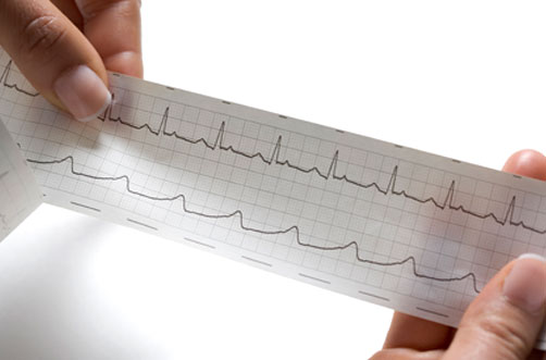
The ECG aspect was referred to as the shape of the ST segment elevation.
Major:
In the dome (type 1), it corresponds to the typical aspect: on the derivation V1, the QRS ends with an elevation of the point J, followed by a superscaled ST segment, with a descending slope sometimes convex upwards , and then a negative T-wave.
Minor:
In the saddle (type 2 and 3), it corresponds to a less typical aspect, with an elevation of the point J, followed by an elevation of the segment ST of concave shape upwards and then of a positive T wave. These anomalies are also found in right precordial derivations. It is not uncommon for such an appearance to follow the typical appearance at several interval ECGs.
Differential diagnosis:
It is a question of differentiating an ECG aspect of Br and an early repolarization syndrome (SRP). The distinction between early athlete heart repolarization syndrome and Br syndrome is not well defined.
The presence of an ST segment elevation in a young trained athlete may lead to a suspicion of Br syndrome.
The aspect of the ECG:
Recently, the ECG aspect of early repolarization of trained athletes was compared to that of patients with Br syndrome in order to identify possible criteria for differentiating the two diagnoses.
The maximum amplitude of the ST segment was measured at point J (ST J) and after 80 ms (ST 80). Finally, an ST J / ST 80 ratio was calculated. An elevation of the ST segment in right precordials was found in 4% of athletes.
Despite similar maximum ST-segment degrees (3.1 ± 0.9 mm versus 3.2 ± 0.6 mm), athletes had on average a lower ST J / ST 80 ratio (0.7 ± 0.13), compared to patients with Br syndrome (1.6 ± 0.3). A ST J / ST 80 <1 had a sensitivity of 87% and a specificity of 100% to identify the athletes. In addition, subjects with Br had a higher heart rate (75 / min ± 9 vs 50 ± 8), a shorter corrected QT (0.35 ± 0.04 s vs 0.39 ± 0.02 s) and a longer QRS duration (0.11 ± 0.2 s vs 0.09 ± 0.01 s).
Thus, the ST J / ST 80 ratio and the duration of the QRS could differentiate the two syndromes.
Bianco (8) analyzes and compares the ECGs of 155 top athletes (83 runners, 38 footballers and 34 cyclists) with those of 50 normal subjects and 25 Br. ECGs. Out of 155 athletes, 139 had an SRP, compared with 18/50 of the normal subjects, with a left precordial localization in 2/3 of the cases.
In an athlete, the SRP was located on the right and convex upwards, identical to the Br. Differences between the two syndromes involved an ST segment elevation, more marked in the Br subjects (4.4 ± 0.3 mm) compared to athletes (2.3 ± 0.6), and duration of QRS (0.11 ± 0.02 s vs 0.090 ± 0.011 s). Bianco suggests distinguishing Br syndrome from the athlete’s SRP by the association of QRS elongation greater than 11/100 e and the presence of an ST overdose of more than 2 mm with a positive predictive value of 100 % and a negative predictive value of 80%.
The stress test:
The stress test does not differentiate the two syndromes.
In the Br, the ST elevation may decrease or even disappear with normalization of the ECG. Anomalies reappear at the recovery phase.
There is no reported arrhythmia.
In the sportsman, the anomalies of repolarization standardize also to the effort.
Finally, in case of doubt, a pharmacological test can be performed. It consists of intravenous injection of 1 mg / kg of ajmalin or 2 mg / kg of flecainide in order to unmask the abnormality. This test should be performed under continuous ECG monitoring and under appropriate safety conditions, since severe ventricular rhythm disorders have been described during its realization.