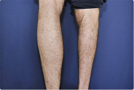
 Muscular diseases are conditions secondary to the pathology of the muscle or the neuromuscular junction. Diagnosis is based on electrophysiological exploration, biology and histological examination of the muscle. Some genes involved are currently identified, with the possibility of a genetic diagnosis.
CLINIC:
Myogenic syndrome:
D IMINUTION OF MUSCLE FORCE:
Proximal especially in myopathic processes, more rarely distal and more or less symmetrical.
– Muscle deficiency of pelvic girdle, trunk and lower extremity root : disorders of the static: hyperlordosis, rocking of the pelvis in front and thorax thrown back, disorders of the walk, waddling with each step inclination of the body towards the supporting member, sign of raising (the patient takes support with his hands on the knees to straighten).
– Deficiency of the muscles of the scapulohumeral belt : decrease in strength (large serratus, deltoid, trapezius, pectoral and biceps) which explains the fall of the stump of the shoulder and the detachment of the shoulder blades.
– Achievement of facial muscles : facies not very expressive (erasure of wrinkles, eversion of lips, transverse smile).The difficulty in closing the eyelids is a sign of the beginning, as well as the difficulty to inflate the cheeks or to whistle.
MODIFICATIONS OF MUSCLE VOLUME:
Atrophy (variable size and extent), sometimes masked by subcutaneous adipose. Muscular hypertrophy is rare.
ABNORMAL MUSCULAR REMOVAL AND CONTRACTION:
The difficulty in relaxation is characteristic of myotonia. Pseudomyotonia is a slowness to contraction and muscle relaxation and is found in hypothyroidism.
A BOLITION OF THE IDIOMUSCULAR REFLEX:
It affirms the myogenic nature of a deficit.
MUSCLE AND TENDER RETRACTIONS:
They are common during certain myopathy.
Negative signs indicate the integrity of the nervous system (preservation of osteotendinous reflexes, absence of sensory or pyramidal signs).
In infants and small children, a muscular pathology will be suspected in front of a weakness of the cry, a difficulty at the feeding, a delay in the motor acquisitions.
Myasthenic syndrome:
– Muscular fatigue during repeated or sustained efforts.
– Deficit variable in time and from one muscle group to another.
– Variable topography. Ocular muscle involvement is almost constant and sometimes the only manifestation.
The ptôsis is very evocative, accentuates with fatigue. Partial ophthalmoplegia results in intermittent diplopia, but intrinsic motility is maintained. Swallowing, phonation and chewing disorders are common. To the members, the motor deficit predominates on the muscles of the roots (difficulty to climb the stairs). The involvement of the respiratory muscles results in dyspnea.
– Mary Walker’s reaction, evocative but inconstant. The repeated contraction of the muscles of the hand and of the forearm, a blood circulation stopped by a tourniquet, causes the exhaustion of the muscles and, at the raising of the withers, we observe a reverberation at a distance, such as the accentuation of a ptosis.
ADDITIONAL EXAMENS:
EMG:
– Specified the mechanism of the injury: myogenic or neuromuscular junction (requires specific techniques: repetitive stimulation or single fiber study).
– Excludes neurogenic damage.
Biological examinations:
– Muscle enzymes (CPK and aldolase).
– A suspicion of pathology of the neuromuscular junction must make search for anti-receptor antibodies of acetyl choline.
– Finally, in case of suspicion of a genetic disorder, a search for the gene involved is sometimes possible.
Others:
– Radiological:
• muscular scanner in case of suspicion of dystrophic pathology;
• thoracic scanner (search for a thymic process if myasthenia).
– Histological: muscle biopsy.
– Stress test with biochemical assay in case of metabolic myopathy.
TECHNOLOGY:
Muscular pathology:
MUSCULAR DYSTROPHY:
Myopathy of hereditary origin. The notion of an identical disease in the siblings is evocative.
Clinical diagnosis, biopsy data, biochemical assays and sometimes evidence of the gene abnormality.
Dystrophy without myotonia:
X-linked
– Duchenne myopathy: it is the most frequent and the most serious (recessive transmission); it begins early in the boy, causes gait disorders and evolves in ten years, deaths by cardiac and respiratory complications;
– Becker’s disease: a variant of Duchenne’s disease, it begins much later in adults and its evolution is much less severe;
– Emery-Dreifuss’ dystrophy: of recessive transmission, it is characterized by the early onset of muscle retractions and disorders of cardiac conduction.
Autosomal dominant transmission:
It affects both sexes:
– facio-scapulo-humeral myopathy: it begins in early childhood. Incomplete occlusion of the eyelids during sleep is the early sign. The myopathic facies and the involvement of the scapulohumeral belt summarize the symptomatology;anomaly on chromosome 4;
– ocular myopathy: impairment of the extrinsic motility of the eyeballs; they begin in their thirty with a ptosis, then an oculomotor attack;
– oculopharyngeal dystrophy: it combines dysphagia and extrinsic opthtalmoplegia. The gene in question is located on chromosome14;
– myopathy of the belts: the deficit can begin at the level of the scapular and / or pelvic girdle with gradual aggravation; heterogeneous group of conditions whose diagnosis is based on histological and biochemical data.
Autosomal recessive inheritance:
Myopathy belts of later onset.
Dystrophy with myotonia
Autosomal dominant transmission:
– Steinert’s dystrophy: myotonia and predominant atrophy on the muscles of the face and neck + distal involvement of the limbs (atrophy in the cuff); early baldness, cataracts, endocrine disorders and cardiac arrhythmias. Gene on chromosome 19. Another form has recently been identified: proximal deficit and myotonia (gene on chromosome 3).
– Thomsen’s disease: myotonia and muscular hypertrophy.
CONGENITAL MOSOPATHY:
Early onset, moderate muscle deficit with hypotonia; very slow or non-progressive evolution. The diagnosis is based on the muscle biopsy.
METABOLIC MOPHATHY:
Clinical tables very variable; semeiology punctuated by the effort with cramps. It may be an abnormality of glycogen metabolism, lipid metabolism or a mitochondrial abnormality.
Diagnosis is based on muscle biopsy, exercise test results and biochemical assays.
MYOSITES:
Polymyositis and dermatomyositis:
Inflammatory disorders associating a predominant deficit at the root of the limbs, muscular pains and cutaneous lesions of the face and extremity. There is an inflammatory syndrome and an increase in muscle enzymes, the diagnosis is confirmed by the histological study. These polymyosite may be secondary to other conditions (malignant tumors, collagenosis).
Infectious myositis:
Trichinosis, cysticercosis, toxoplasmosis, toxocariasis or viral diseases (HIV, coxsackie).
Myositis with inclusions:
It occurs after age 50: progressive muscular deficit, painless overall or predominantly distal. The diagnosis is based on the muscle biopsy.
TOXIC MOPHATHY:
Lipid-lowering agents (fibrates or statin), chloroquine, fluorinated steroids (high doses, prolonged duration), antiretrovirals (zidovudine, azidothymidine).
ENDOCRINE AND NUTRITIONAL MOSOPATHY:
Thyroid myopathy (hyper- or hypothyroidism), more rare, acromegaly, Cushing, Addison, hyperparathyroidism.
The ethylism can give an acute myopathy and a chronic form. Osteomalacia is a rare cause.
PERIODIC PARALYSY:
Hypotonic and muscular weakness of variable duration, extent and intensity associated with changes in serum potassium; autosomal dominant transmission disorders (mutations in the sodium channel gene some of which are amenable to genetic diagnosis).
Pathology of the neuromuscular junction:
MYASTHENIA:
Autoimmune pathology (anti-acetylcholine receptor antibody); abnormal fatigability on exertion, secondary to a post-synaptic neuromuscular transmission disorder. Diagnosis is based on electrophysiological exploration, anti-receptor antibody research and anticholinesterase enhancement (prostigmine test). The search for a thymic anomaly (thymoma or hyperplastic remnants) is systematic. Many drugs are contraindicated in case of myasthenia gravis (curare, aminoglycoside, colimycin, erythromycin, betablockers, phenytoin, dantrolein, quinine, procainamide, phenothiazine, benzodiazepine or lithium).
SECONDARY MYASTHENIC SYNDROME:
With the administration of D penicillamine, the bite of certain snakes, poisoning with combat gases …
MYASTHENIC SYNDROME BY NEUROMUSCULAR BLOCK OF PRESYNAPTIC TYPE:
– Botulism: occurs 12 to 36 hours after ingestion of the toxin; nausea and vomiting, visual disturbances, diplopia, dry mouth, proximal motor deficit, mydriasis, intrinsic and extrinsic ocular motility disorders, facial and pharyngeal motricity.The respiratory muscles can be reached in severe forms.
– Lambert Eaton Syndrome: occurs during the course of cancer (small cell lung carcinoma), but can sometimes be autoimmune. Fatigability with relative increase in muscle strength during repeated contraction; dryness of the mouth with metallic taste in more than 50% of the cases, motor deficit especially net on the muscles of the pelvic girdle and the thighs; osteotendinous reflexes are very often abolished in the lower limbs.

Leave a Reply