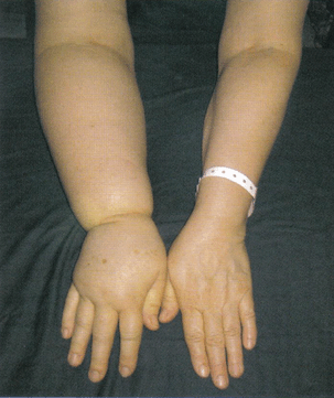
The “big guys” is the colloquial term corresponding to the upper extremity lymphedema. It is a disease whose origin is almost exclusively secondary treatment of breast cancer although there are other rarer causes (axillary lymph node biopsy for diagnostic purposes, Hodgkin and non-Hodgkin lymphoma treated with axillary irradiation). [1]
Prolonged lymphatic stasis increases the volume of the affected limb.
This chronic disease can be disabling the functional, aesthetic and psychological and cause an impact on professional, relational and social.

DEFINITION AND EPIDEMIOLOGY:
Lymphedema of the upper limb secondary to breast cancer is the leading cause of lymphedema in France. The frequency is currently estimated at between 15 and 28% after conventional axillary dissection and between 2.5 and 6.9% after sentinel lymph node biopsy.
The variability of the percentages can be explained by variables observation times and definitions of variables lymphedema, comparing the affected limb and contralateral limb: perimeter differences of 2 cm or volume of 200-250 mL.
TIME FOR APPEARANCE:
Lymphedema can appear immediately after surgery or much later, until 20 years after treatment. The median time of onset is 2 years.The natural course of lymphedema is toward the volumetric increase with aggravation of the tissue component (adipose tissue, “fibrosis”). In very rare cases, lymphedema of the upper limb may be indicative of breast cancer.
DEVELOPMENT OF A RISK FACTORS LYMPHEDEMA:
The main risk factors for developing lymphedema are the number of nodes removed during axillary dissection, radiation therapy, especially on axillary lymph node areas, mastectomy (lumpectomy vs), overweight when breast cancer, weight gain after cancer treatment and reduction of activity after breast surgery. Furthermore, the severity of lymphedema is correlated with the Body Mass Index.
CLINICAL EXAMINATION:
Heaviness, pain:
The impression of heaviness is the most common symptom, sometimes described as a feeling of heaviness in the affected limb lymphedema. The pain is much less common, and should suggest an associated plexopathy or post-radiation or by tumor invasion, toxic neuropathy, deep vein thrombosis, a condition of the shoulder (with limited mobility) or a syndrome carpal tunnel.
Skin and appendages:
The skin may be flexible or otherwise tense (not pleatable), take the bucket, or have a look elephantiasis.
Assessment of the volume of lymphedema:
This is an essential step that is practiced before / after treatment and at follow-up of lymphedema.
The reference technique is the water volume, but in practice, we use the perimeter measures taken at regular intervals (every 5 or 10 cm) which calculate a volume in mL by assimilation of members of segments of truncated cones .
TESTING:
No exploration is required in the diagnosis of lymphedema. However, a venous Doppler is useful when the onset or worsening of lymphedema. In case of tumor recurrence is suspected, a CT scan or MRI of the axilla or a PET scan is needed.
COMPLICATIONS:
The main complication of lymphedema is erysipelas. Most of the time, we did not find any infectious door. Treatment is with antibiotics (amoxicillin, pristinamycin). Sometimes recurrences are common and require prolonged antibiotic whose terms are not consensual. Osteoarticular complications of the shoulder can occur when the volume of the member is important. The occurrence of lymphangiosarcoma (or Stewart Treves syndrome) is very rare and poor prognosis.

Figure 1. Bandage with little elastic bands Somos® a NN® foam padding to secondary lymphedema of the upper limb.
TREATMENT:
The treatment of lymphedema based on the prevention of infections (erysipelas) avoiding risk procedures (cuts, scrapes, burns).
The other side of the management is to reduce the volume of lymphedema. The complete decongestive physiotherapy treatment called is divided into two phases: the first intensive is designed to reduce the volume and high maintenance to maintain the reduced volume.
The principal element is the daily application, 24h / 24h, little elastic bandages with strips short elongation (<100%), (Somos®) (Fig. 1) on a foam padding and / or cotton, possibly preceded by manual lymph drainage. The duration of the intensive treatment is 1 to 3 weeks.

Figure 2. elastic compression sleeve taking the hand for secondary lymphedema of the upper limb after breast cancer.
During the maintenance phase, after obtaining a reduction in volume, the port, the day of a class of elastic compression sleeve of 3 or 4 is essential. [2] It is best to prescribe a sleeve taking the hand (with attached mitten) to prevent it swells (Fig. 2). Their wear frequently replaced every 3 to 4 months. The realization of inelastic bandages at night by the patient himself after learning with a physical therapist at a frequency lower than that of intensive treatment (3 per week) is associated with daytime port sleeve. Skin Care with regular moisturizing night is often necessary. If overweight, weight loss can promote the reduction in volume of lymphedema. The veinotonic drugs are very inefficient and banned diuretics.

Leave a Reply