DIAGNOSIS:
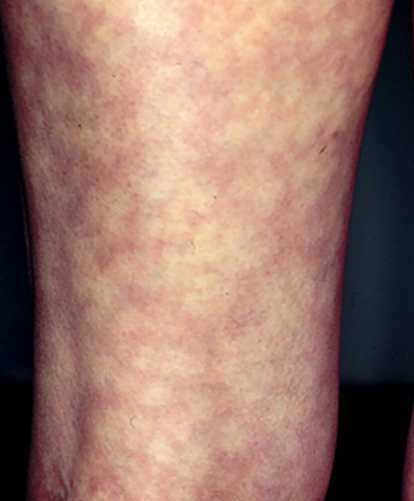
The clinical appearance of livedos is the purple mesh on the skin.
Livedo corresponds to a blood stasis in the superficial venous plexus of the dermis.
The diagnosis of livedo poses no problem. The main difficulty is the etiologic diagnosis. Evident in livedos due to low circulation rates, it can be a serious systemic illness.

Clinical examination:
We distinguish clinically by their appearance the branched and crosslinked livedos.
Branched or livedo racemosa:
The meshes are large, not closed, irregular and asymmetrical.Livedo may sit on the buttocks, thighs, arms, trunk.
It does not completely disappear in supine and warming.
A systemic cause should be investigated.
Crosslinked Livedo:
The meshes are smaller sizes closed.
Livedo appears during orthostatic, cold, and disappears supine and warming.
It is most often physiological, particularly in infants; a general cause is rare.
If livedo branched general cause is suspected, a skin biopsy may be performed at the center of the mesh to identify the involved vascular abnormality (arteriolar damage, blood viscosity).
Differential diagnosis:
Some skin diseases can mimic livedo as the application of heaters or other sources of prolonged heat.
These can lead to crosslinked pigmentation not attenuating the heat and vitropression. They are most noticeable on the anterior surface of the legs and thighs.
Treatment is based on the eviction of the triggering cause.
ETIOLOGY:
Vasomotor disorders:
Livedo is crosslinked. The physiological causes are the most common, especially in infants and children. Low circulation rates are the other main cause.
The crosslinked livedo is rarely seen in carcinoid and pheochromocytoma. Amantadine, used in Parkinson’s disease, may also lead livedo.
Livedo branched:
Thrombosis:
Thrombosis is a cause of livedo branched. This is seen especially in the antiphospholipid syndrome, primary or secondary to connective (especially lupus). In livedo branched may be associated Raynaud’s phenomenon, acrocyanosis, distal necrotic lesions, subungual splinter haemorrhages, leg ulcers, superficial or deep thrombophlebitis signs.
Treatment is based on the longer extended anticoagulation (INR around 3) associated with that of the underlying disease.
A coagulation profile is also necessary (search for personal or family history of thrombosis and miscarriage, dosage of antithrombin, protein C and S, search for the G20210A factor II and a resistance to protein activated C).
Embolism:
We must think of cholesterol embolism occurring before a livedo, the waning of an arterial gesture in a subject atheromatous anticoagulants.
Purple toes are a classic sign. The fundus and skin biopsy may reveal emboli. A gradual and delayed renal failure, fever, neurological dysfunction or an aspect of pseudopériartérite wrack (PAN) can be seen.
Treatment is based on the eviction of risk procedures in these patients, the cessation of anticoagulant and sometimes systemic corticosteroids.
The prognosis is not good.
More rarely, we will discuss:
– A myxoma of the left atrium: the diagnosis is made by echocardiography.
Treatment is based on surgery and anticoagulation;
– A marasmus endocarditis in a subject with advanced cancer, with multiple possible emboli. The prognosis is terrible;
– Cancers (pancreatic cancer, lymphoma angiotrope);
– Viral infections rarely cause a branched livedo, more by qu’embolique vasculitic mechanism (CMV).
Systemic diseases:
This can be observed in livedo vasculitis
systemic, relapsing polychondritis, etc.
Sneddon Syndrome:
Sneddon syndrome is defi ned by the association of a branched livedo extended and permanent ischemic stroke.
Some of these livedos match the antiphospholipid syndrome (APS). Moyamoya syndrome can approach.
The patient is entrusted to the specialist.
Anticoagulation is required.
Hyperviscosity:
Hyperviscosity is seen in myeloproliferative disorders and monoclonal gammopathies high rates. In livedo may associate acrocyanosis, erythermalgia, érythrosique facies, headache, ringing in the ears, blurred vision, etc.
Treatment is symptomatic and etiologic (hemodilution, bled, antiplatelet agents, etc.)
Metabolic:
Sometimes necrotic livedos observed in secondary hyperparathyroidism in chronic renal failure dialyzed in hyperoxaluria, etc. These calcium crystal deposition or oxalate.
The treatment is disappointing, correction of metabolic disorder not involving the disappearance of the lesions.
Medications:
Include quinine, beta-blockers, amantadine, phenylbutazone. Livedo is often crosslinked. The Livedoid dermatitis Nicolau, due to the intra-arterial injection of medicines accidentally, associates with livedo pain after injection with risk of secondary necrosis.
Hospitalization is required for medical treatment (vasodilators, heparin) and surgical excision of necrotic lesions.
Leave a Reply