
Telangiectasia are expansions of the superficial dermis vessels. If they are usually benign, they may nevertheless reveal a general disease that will require thinking, scleroderma (particularly the form of CREST [subcutaneous calcinosis, Raynaud’s phenomenon, esophageal dysfunction, sclerodactyly, telangiectasia] ), Rendu-Osler disease primarily;Exceptionally, the ataxietélangiectasie and a particular form of cutaneous mastocytosis (Telangiectatica eruptiva macular perstans).
DIAGNOSIS:
The patient often consults for disfigurement. The diagnosis is clinical.
The main clinical features are:
– Pink or red spot, edged, square, without vascular arborization (macular telangiectasia). These forms are encountered in CREST and Rendu-Osler disease;
– Hair Scalp: Linear trail or network, pink or blue, without relief. This is the aspect observed on the lower limbs in case of venous insufficiency;
– Small papule dew edged. This is the aspect of the ruby stain (or senile angioma)
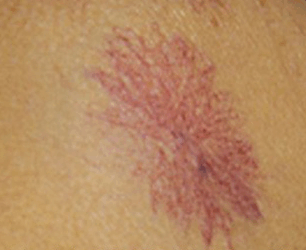
– Spider angioma: a red central point leave centrifugal arborizations, disappearing to the pressure on the central point. It occurs mostly in cases of liver failure and during pregnancy.
Erythema telangiectasia disappears pressure, unlike purpura. The surface is not keratinocyte, unlike angiokeratoma.
ETIOLOGY:
The clinical appearance of the skin lesion, terrain, location and association with other symptoms used to guide the diagnosis.
Macular telangiectasia, quadrangular, edged:
It is then necessary to refer to two causes: an acquired, systemic sclerosis (mainly in its limited form, CREST syndrome) and hereditary (Rendu-Osler disease).
Scleroderma:
This mainly CREST syndrome with subcutaneous calcinosis, Raynaud’s syndrome, esophageal reached, sclerodactyly and telangiectasia.
Cutaneous sclerosis is predominantly acral, reaching the face and neck, but not the trunk or root members. The facial appearance is characteristic, with a topspin aspect, a bird beak profile, limitation of mouth opening.
Telangiectasia quadrangular are also seen on the palms. Sclerodactyly is clear. To these partners Raynaud’s syndrome often before the onset of other signs and can be complicated by digital ulcers and trophic disorders.
Capillaroscopy found signs of microangiopathy characteristics. Gastroesophageal reflux with esophagitis can often disabling and can be complicated by Barrett’s esophagus.
Subcutaneous calcinosis is sometimes disabling, relapsing inflammatory and whitish material outcome. Power structures of the skin can worsen trophic disorders.
The particular signs search examination for pulmonary arterial hypertension (chest pain, faintness, and dyspnea on exertion appears, Signs rights, B2 burst of pulmonary fireplace) and a rare pulmonary fibrosis in these forms Scleroderma (bilateral crackles, purposes and dry predominant bases).
Laboratory tests to be performed are limited: creatinine, NFS (complete blood count), looking for centromere antibodies in localized forms and anti-Scl 70 in diffuse.
The patient is told the hospital specialist.
Despite recent therapeutic advances, there is no satisfactory treatment of this background sometimes dreaded disease.
Hereditary hemorrhagic telangiectasia:
The Rendu-Osler disease can be from childhood by epistaxis.
Telangiectasia, to initially look similar to scleroderma, appear thereafter.
They can take relief in the course of evolution. Telangiectasias are found on the face, hands where they predominate, but also on the mucous membranes (lips, tongue).
Capillaroscopy shows abnormalities in the majority of cases.
The complications are mainly bucconasales repeated bleeding, which can lead to anemia, and visceral (fibrosing liver injury possibly associated with arteriovenous fistulas, pulmonary arteriovenous fistulas that can cause PAH [Pulmonary Hypertension] and cerebral abscess).
Mark family history is important because it is a genetic disease, possibly linked to a mutation of the TGF-β gene.
Treatment is purely symptomatic. The patient is assigned to a specialist to support any visceral complications.
Ataxia telangiectasia:
Telangiectasia are here mostly type scalp hair appearing around the age of 3 to 5 years. They affect the conjunctiva, nose, ears, bend the elbows. Appear secondarily cerebellar syndrome and recurrent infections associated with immunosuppression, photosensitivity with possible skin cancers.
The management is done by specialized teams.
Telangiectasia macularis eruptiva perstans:
This is a rare form of mastocytosis with rare systemic involvement. This is erythematous macules and papules or pigmented dotted with telangiectasia.
Various causes:
Telangiectasia may also occur in dermatomyositis, lupus (pseudocouperosique appearance).
Telangiectasia located:
Face:
Apart from the cases already described, we can mention: spider nevi: liver disease with liver failure, mostly found during pregnancy and in children;
– Association flushing, particularly triggered by hot, alcohol, emotion or pustules: think of rosacea.
_ On the upper chest
There may be physiological telangiectasia type of arborizations blue. They should not be confused with chest collateral venous circulation of the superior vena cava syndrome. Recall the chest location of the telangiectasia macularis eruptiva perstans.
The lower limbs:
The ankles, calves, thighs, we must think of venous insufficiency, especially if it is associated with a Vesper edema in the middle-aged woman. Personal history of venous thrombosis and family liabilities of venous insufficiency are sought.
A Doppler ultrasound of the lower limbs in search of venous insufficiency is helpful.
The assumption is based mainly on the venous compression (often mean by venous compression stockings, veinotonic treatment, sclerotherapy).
Ankles:
There may also be hair hairy ankles in women is gradually extending upward and can win the abdomen. There may be a family history.
There is no treatment.
Other causes:
Viral infections:
Telangiectasia acquired can be observed in viral infections, especially echovirus.
There is often a vasoconstriction ring around papular telangiectasia.
Spontaneous regression is the rule.
Telangiectasias of acquired also have been described in HIV subject, type of scalp hair and a rash with periungual telangiectasia also among those infected with HIV and HCV (hepatitis C).
Toxic taking:
Some drugs have been implicated in the occurrence of spider veins, such as calcium channel blockers, with photo-induced telangiectasia on the face and chest.
Some toxic have been implicated (aluminum).
In prolonged corticosteroid therapy may be associated telangiectasias, accompanying skin atrophy, purpura Bateman and Cushingoid appearance.
Other:
Very rare cases of angiotrope lymphoma have been described.
The serpiginous angioma Hutchinson is a beginner hemangioma in childhood as a plan angioma. Appear secondarily peppercorn in telangiectasia with a rough surface to the touch. Angioma covers the legs and buttocks serpiginous manner. The condition is benign and can regress spontaneously.
Telangiectasia are also found in Cushing’s syndrome.
TREATMENT:
Besides the already mentioned treatments, treatment of telangiectasia itself is symptomatic.
The lower limbs, sclerotherapy has been proposed.
Vascular lasers can be of great help, as well as topical retinoids.
In case of general illness associated support telangiectasias, and their possible complications premium.
Leave a Reply