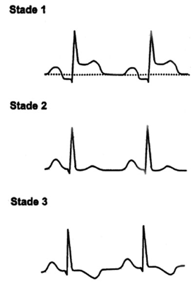
Developments in 4 stages:
Stage I:
Subepicardial injury: ST segment elevation in upper concavity, early, fleeting, not including the T wave, positive.
Stage II (2nd week):
ST return to the isoelectric line with flattened T waves
Stage III:
Negativity of the T wave, which can persist for several weeks (most constant sign and most durable: 2-6 weeks)

– Essential fact: it’s images are diffuse and no images in mirrors.
– Other signs: QRS low volts (microvoltage) if effusion; sub-offset PQ segment (of high diagnostic value);extrasystoles and atrial fibrillation are common; appearance of alternating electrical abundant effusion (changes the direction of the electrical axis of the heart).
– Tamponade: the only sign of value is the electrical wave (variation of the amplitude and the morphology of the QRS, with variation of the electrical axis of the heart).

Leave a Reply