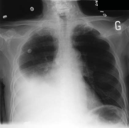
 This frequent symptom is observed in many clinical pictures. The diagnosis of pleurisy is clinical and a simple radiograph of face and profile is usually sufficient to confirm the diagnosis.
The pleural puncture is most of the time essential to the research of the etiological diagnosis.
This is often easy with the context but can be difficult, when it is an isolated symptom and when several causes combine to cause a serous effusion.
The clinical examination is the essential time when the diagnosis is not obvious; we must look for antecedents and especially the notion of tuberculous contagion, the existence of cardiopathy, the introduction of a new treatment. An alteration of the general condition or the existence of a fever must be noted. The physical examination must specify the existence of cardiac insufficiency, sloping abdominal dullness, organomegaly, peripheral lymphadenopathy …
ADDITIONAL EXAMINATION:
The pleural puncture is most of the time essential to the search of the etiological diagnosis except if the cause is certain (heart failure) or if the quantity of liquid is too little abundant.
Examination of the liquid:
BIOCHEMICAL ANALYSIS:
Differentiating exudate and transudate is essential because if the fluid is a transudate, it is not necessary to perform costly and invasive supplemental examinations. The clinical examination will most often determine the cause: heart failure especially.
In principle, and always correlating the clinic, a protein level> 30-35 g / l is sufficient to distinguish exudative pleurisy.The use of this criterion alone, however, may result in false classifications, particularly if the patient is on diuretics.Other dosages and criteria will be studied in difficult cases (determination of blood and pleural LDH, or albumin).
CYTOLOGICAL EXAMINATION:
A lymphocyte fluid evokes tuberculosis, but some lymphocyte serities are due to lymphomas, the distinction between reactive and malignant lymphocytes being sometimes difficult in the morphological analysis alone. Transudative fluids, heart failure, cirrhosis and nephrotic syndrome are poorly cellular (mesothelial cells, mononucleated cells).
The presence of eosinophilia> 10% is rare. Eryosinophilia evokes a malignant or toxic effusion (induced by a drug).Asbestosis, Churg’s disease and Strauss can produce eosinophil-rich effusions as well as tuberculosis.
A predominance of polymorphonuclear is mainly due to bacterial pleurisy.
The search for malignant cells in serous fluids is a usual concern because the appearance of an effusion is frequent in the evolution of a cancer. The metastases of a mammary adenocarcinoma are the most common cause in women, while lung cancer and malignant mesothelioma account for a relatively large number of cases in both sexes.
BACTERIOLOGICAL ANALYSIS:
Any liquid collected must be sent in bacteriology. Direct examination after Gram staining should be performed, as well as culturing in search of the usual pyogenic and anaerobic germs.
The diagnosis of tuberculous polyseritis is one of the most difficult diagnoses. The direct examination is insensitive, the culture more sensitive but still a little long. The sensitivity of PCR is variable.
Other complementary examination:
Standard radiological examinations: chest X-ray, thoracic computed tomography, are essential for the etiological diagnosis to direct further investigations.
The biopsies, possibly ultrasound-guided or CT-guided or, if necessary, directed under thoracoscopy, have a better diagnostic sensitivity in the malignant or tuberculous serities than the cytological examinations of the liquid or the blind biopsies.
CAUSES OF A PLEURAL EFFUSION:
Serous transudative effusions (proteins <30 g / L):
They are in principle of easy diagnosis on the clinic, the standard biology and the simple radiology and do not require the recourse to the other complementary examinations.
Etiologies of a transudative serous effusion:
Etiologiy:
Cardiovascular:
Heart failure
Chronic pericarditis constrictive
Pulmonary embolism
Obstruction of the VCS
Hepatic and digestive:
Cirrhosis
Hepatocellular insufficiency
Acute alcoholic hepatitis
Exudative enteropathy
Renal:
Nephrotic syndrome
Peritoneal dialysis
Others:
undernutrition
Hypothyroidism
sarcoidosis
Demons-Meigs Syndrome
Congestive heart failure is the most common cause of transudates, in bilateral rule.
Cirrhosis is responsible for sometimes abundant effusions, usually always accompanied by clinical ascites. Other causes are rarer.
Exudative effusions (proteins> 30 g / L):
Three main categories of causes predominate, cancers and hemopathies, tuberculosis and inflammatory causes. The pleurisy is rarely isolated and the clinical context, of course, very contributory to the diagnosis
Etiology of an exudative effusion:
Cancers:
lungs
breast
Digestive tract
Pancreas
Ovary
mesothelioma
lymphomas
Infections:
parapneumonic
Tuberculosis
Inflammatories:
Lupus
Rheumatoid arthritis
Familial Mediterranean fever
Still’s disease
sarcoidosis
Infectious pneumonias are very frequent, bacterial (pneumococcal, intracellular germs) or viral (often hyperalgic). The context most often allows the diagnosis.
Tuberculous pleurisy remains present in our countries, migrant, immunocompromised populations.
Metastatic pleurisy is the most common exudative pleurisy after 50 years, the diagnosis of primitive is sometimes difficult when the cancer is not known and the use of directed biopsy is often necessary, this is also the case of mesothelioma. The pleurisy of lymphopathies is also very frequent. Other causes are rarer.
Some medicines are likely to cause, much more rarely pleurisy: Clozapine,
Taxanes, Ergot Derivatives, Praziquantel, Methotrexate, Ramipril, Cordarone, Carbamazepine.
We must add rare causes:
– pleurisies of sub-diaphragmatic pathologies, pancreatitis, abscesses, whose diagnosis is in principle obvious;
– exudates of cardiovascular, postembolic origin (which may cause diagnostic problems), postinfarction;
– hypothyroidism, usually profound and obvious diagnosis, pleurisy is then often accompanied by indolent pericarditis.
SPECIAL CASE OF POLYSTERY:
In front of a polyserite, when the diagnosis does not seem obvious, one must surely make maximum use of the biochemical, cytological and bacteriological results taking care to confront them to the history of the patient, the epidemiological context and the clinical signs.
– Always carry out a diagnostic sample. It is always best to take a sample of serum at the same time and send them together to the laboratory.
– Take note of the macroscopic appearance of the liquid.
– Distinguish between transudate and exudate in pleural effusions and, in the case of ascites, confirm the presence or absence of portal hypertension by GASE.
– If the effusion is a transudate, the other tests are of no use, often giving misleading information. The clinician should state or rule out the usual causes such as congestive heart failure, hepatic cirrhosis, renal failure and start treatment.
– If the effusion is an exudate, additional examinations will be necessary and will depend on:
• the macroscopic appearance of the liquid;
• the clinical picture;
• origin of the patient: tuberculous effusions are common in developing countries. ADA can be useful in these areas of high tuberculosis prevalence, providing a rapid and useful diagnostic orientation, thus speeding up initial decision-making.
The etiologies of polyserites are multiple, but they are still dominated by cancers, hematological diseases, tuberculosis and collagenosis.
Leave a Reply