* Pleural opacities have an obtuse angle connection with the chest wall in contrast to parenchymal opacities juxtapleurales that connect to an acute angle.
* Opacities mediastinal their internal boundary coincides with the mediastinum and outer boundary connects gently sloping with it.
* Multiple round opacities (drop ball) are usually metastases.
* A doubling within 7 days or no doubling over 2 years are arguments benignity.
* As the diameter increases further the risk of malignancy is high. 80% of benign nodules are less than 2 cm.
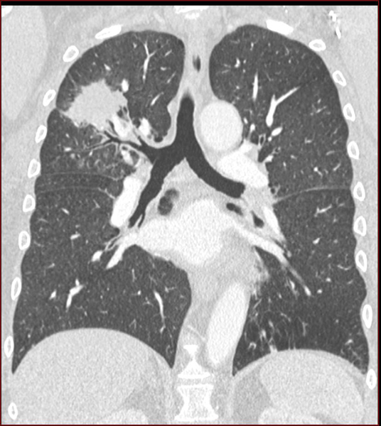
* 70% to 95% of neoplastic nodules have fuzzy boundaries and spiculated.
The lobulated contours are also argue malignancy.
Conversely the presence of micronodules outskirts of a round opacity oriented more towards TB. Calcifications are benign argument.
The character excavated is not seen in tuberculosis and may be tumor.
Air bronchogramme does not always mean an infectious and can be reached in case of bronchoalveolar carcinoma or lymphoma.
* The presence of lesions extraparenchymateuses: bone resorption, pleural effusion, mediastinal ADP are very suggestive of neoplasia.
* Arguments biological (after clinical and radiological orientation): smear; Research Aspergillus in sputum; hydatid serology and aspergillus …
* Bronchial Fibroscopy: aspirations and bronchial brushing allow a cytological and bacteriological study. This examination is essential to pretherapeutic round opacities.
* Suspicion of infectious causes (tuberculoma, aspergilloma, hydatid disease) and vascular (anévérismes …) cons-indicated biopsies (risk of infectious spread, bleeding risk).
* For other cases -> definitive diagnosis: histological sampling.
Bronchoalveolar lavage and transbronchial biopsies (distal lesions not directly accessible). Transparietal puncture.
* Exploratory thoracotomy: extemporaneous histological examination.
* The pulmonary hamartoma (hamartochondrome) is the most frequent lung tumor (size ≤ 2.5 cm regular contours …)
* Hydatid cyst: homogeneous opacity of 1 to 10 cm in diameter, with particularly sharp and regular outline, rather serving on bases and right of fluid tone without density enhancement on CT after contrast injection.
The presence of the air out of the sign cyst in the bronchi.
The discovery of liver cyst and positive serology assist in the diagnosis.
* Aspergilloma: radiological appearance “in bell” associated with the old radiological lesions. Dosage precipitin Aspergillus …
* Atelectasis by winding (often associated with pleural abnormalities) the arcuate path of the vessels and bronchi entering the round opacity with its lower pole is evocative.
* Pulmonary infarction (moving towards flat atelectasis before disappearing).
* Lung opacities (mucoid impaction)


Leave a Reply